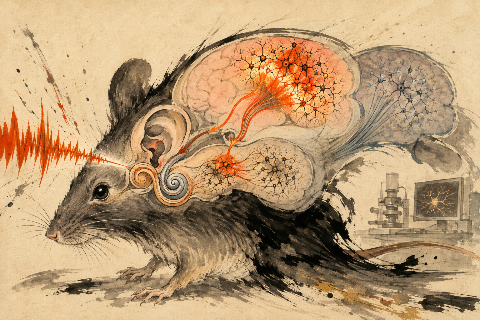
A refrigerator’s hum can disappear from awareness without ever becoming quieter. A glass breaking across a crowded room does the opposite: one instant of acoustic difference cuts through the scene. The ears transmit both. The decisive editing happens farther inside, where the brain suppresses what has become predictable and boosts what violates the pattern.
Researchers led by Shigeo Okabe at the RIKEN Center for Brain Science, working with Shunsuke Mizutani, Yasuhiro Go, Atsu Aiba, Kiyoto Kasai and colleagues at the University of Tokyo, University of Hyogo and Japan’s National Institutes of Natural Sciences, have now resolved part of that editor at cellular scale. Their study appeared in Science Advances on August 14.
The team found the strongest response to a rare, different-pitch tone in the shallow layers of higher-order auditory cortex. The responsive neurons did not lie randomly. They formed local clusters, and the cells whose activity was most synchronized with their neighbors showed the strongest deviance detection. A mouse carrying a deletion corresponding to human chromosome region 22q11.2 showed a selective weakening at each step: fewer deviance-detecting cells, weaker clustering, lower synchrony and a smaller amplified response.
The oddball test: create a rule, then break it
The experiment rests on a remarkably spare design. Play one pitch again and again. Once the sequence has established a regularity, insert a rare tone of a different pitch. The repeated tone is called the standard; the rare one is the deviant. In people, subtracting the brain’s response to the standard from its response to the deviant reveals a characteristic event-related potential called mismatch negativity, or MMN.
But a bigger response to the rare tone can have at least two causes. Neurons may simply tire of the repeated standard, a process called repetition suppression or stimulus-specific adaptation. Or the brain may actively register that the deviant violates an inferred rule. A classical oddball comparison mixes those two effects.
The RIKEN-led team therefore included a control sequence of randomly varying pitches. A tone in that sequence is infrequent without following one dominant, repeated standard. Comparing physically equivalent sounds across contexts helps separate “rare because it was not repeated” from “surprising because it broke a pattern.”
Deviance detection ≈ response to deviant − response to matched random-control tone
Adaptation ≈ response to random-control tone − response to repeated standard
| Sequence | What the mouse hears | What the comparison isolates |
|---|---|---|
| Repeated standard | The same pitch occurs again and again. | Response after a sensory representation has adapted to repetition. |
| Deviant, or “oddball” | A rare different pitch interrupts the repeated tone. | Adaptation plus sensitivity to a broken regularity. |
| Random control | Different pitches appear without one dominant repetition. | A baseline for rarity and physical sound features with less predictable context. |
This subtraction is not philosophical decoration. It changes the biological claim. “The neuron likes a fresh pitch” is weaker than “the neuron responds because this pitch is wrong for the current context.” The study’s cluster-and-synchrony effect tracked the deviance-detection component, not the adaptation component.
Watching hundreds of cells in a living brain
The researchers used two-photon calcium imaging, which sends ultrashort infrared laser pulses into fluorescently labeled tissue. When a neuron becomes active, calcium enters the cell and changes the reporter’s fluorescence. Repeated imaging can follow many identified cells while the animal hears the sound sequences.
Calcium is an indirect measure of electrical activity. It is slower than the millisecond voltage changes that form a human EEG, and converting fluorescence into an exact number of action potentials requires assumptions. Its advantage is spatial: the image shows which cells participated and where they sat, something a scalp electrode cannot do.
The team compared low- and higher-order auditory areas, superficial and deeper cortical layers, and axons bringing input from the thalamus into auditory cortex. This anatomical ladder mattered. If the model’s abnormality already appeared in the incoming thalamic signal or throughout every auditory layer, there would be little reason to single out a local cortical computation.
| Measurement level | Control mice | 22q11.2-deletion model |
|---|---|---|
| Thalamic axons entering auditory cortex | No selective cortical-cluster result originates here. | The RIKEN release reports no significant genotype difference in the measured calcium response. |
| Lower-order auditory cortex | Processes basic acoustic features; no clear deviance-cell cluster was found. | No significant genotype difference in the deviant response reported for this lower stage. |
| Superficial higher-order auditory cortex | The strongest deviant response; detectors cluster locally and synchronize. | The strong response is markedly reduced; detectors are fewer, less clustered and less synchronized. |
| Repetition adaptation | Present, but its strength does not track local synchrony. | The disease-relevant difference centers on deviance detection rather than a blanket loss of adaptation. |
The defect was not “the auditory system is quiet.” Incoming sound responses and lower-order processing were comparatively preserved. What weakened was the cortical multiplication applied to a contextual violation.
A cluster is a team, not a lump
“Clustered” can be misleading. The researchers did not discover a new anatomical organ or a visible knot of cells. They found that neurons classified by their response to deviant sounds were spatially closer to one another than expected in the higher-order area. Their activity was also functionally coupled: the more tightly a cell’s activity moved with surrounding cells, the larger its deviance-detection signal.
The distinction between co-location and connection is important. Cells can sit near one another without sharing strong direct synapses, and they can synchronize because they receive a common input. The paper’s title emphasizes strong connections among locally clustered upper-layer neurons, while the imaging establishes the coordinated population pattern. Mapping the exact synapses and causal direction remains a task for circuit tracing, paired recordings and targeted perturbation.
The idea has a long lineage. In 1949, psychologist Donald Hebb proposed that repeatedly coactive neurons could form a “cell assembly,” a distributed unit capable of carrying a perception or memory. Modern imaging has replaced the metaphor with measurable ensembles: groups defined by correlated activity, shared selectivity and, sometimes, causal influence on behavior. The RIKEN study places auditory deviance detection inside that ensemble tradition.
Why the upper layers are plausible messengers
The cerebral cortex is laminated. Thalamic sensory input is especially prominent in middle layers, while superficial layers 2 and 3 exchange information across cortical areas. Their pyramidal neurons are well positioned to combine incoming acoustic evidence with context from neighboring and higher-order regions, then broadcast a result elsewhere.
That architecture fits predictive coding, a framework in which higher levels convey expectations and lower or intermediate circuits signal the mismatch between expectation and input. The framework is influential, but “prediction error” should not be treated as a substance photographed under the microscope. It is an interpretation supported when a response depends on context, survives controls for repetition and rarity, and moves through the hierarchy in a direction predicted by the model.
Independent primate work sharpened this picture in 2023. A University of Tokyo and RIKEN-affiliated team imaged awake marmosets and found offset responses to deviant tone durations in layer 2/3 of a higher-order region called the rostral parabelt. The signal spread into layer 1 of primary auditory cortex; blocking the higher-order area prevented deviance detection in the primary area, while optogenetic activation enhanced the primary response. The new mouse study asks a complementary question: how is an error-like signal locally organized, and what changes in a disease-relevant genotype?
The molecular clue: Baz1a is a label, not yet a lever
Imaging classifies a cell by what it does. Transcriptomics classifies it by the genes it expresses. To connect the two, the researchers isolated cells from auditory, somatosensory and prefrontal cortex and analyzed gene-expression patterns one cell at a time. Among three upper-layer excitatory populations marked by Adamts2, Agmat or Baz1a, the Baz1a-positive population aligned with the deviance-detecting group. Its proportion was selectively reduced in the deletion model.
That gives the circuit a possible molecular address. It may eventually allow researchers to target the relevant population, compare it across species or ask which developmental programs make its connections unusually strong.
It does not show that loss of Baz1a causes schizophrenia, that Baz1a protein itself computes surprise or that increasing it would repair the circuit. A marker can be the flag on a cell type without being the engine that gives the cell its function. The 22q11.2 deletion also removes a block containing many genes, so the chain from copy-number change to cell identity to synaptic organization is almost certainly multistep.
From a 1978 waveform to a cellular map
Risto Näätänen, A. W. K. Gaillard and Sirkka Mäntysalo’s 1978 paper reinterpreted an early negative brain-potential difference during selective listening. The response became known as mismatch negativity. Unlike the later P300, which is strongly tied to attention and task relevance, MMN can be elicited while a person is not consciously attending to the sounds.
Over the next four decades, researchers showed that MMN responds not only to pitch but also to changes in duration, intensity, location and learned acoustic patterns. Source-localization, intracranial recording and animal studies placed change-sensitive activity at multiple levels of the auditory hierarchy. The simple “standard minus deviant” waveform became a window onto sensory memory, statistical learning and contextual prediction.
1949 — Donald Hebb formalizes the cell-assembly idea: groups of coactive neurons can become functional units.
1978 — Näätänen, Gaillard and Mäntysalo publish the evoked-potential result that anchors the history of MMN.
1990s–2000s — Repeated clinical studies find smaller auditory MMN in schizophrenia, especially in chronic illness.
2009 — A major mechanistic review frames adaptation and model adjustment within predictive coding.
2019 — Human electrocorticography localizes a strong deviance-detection contribution in lateral superior temporal gyrus.
2020 — A University of Tokyo study using a many-standards control finds impaired deviance detection, but preserved adaptation, in schizophrenia.
2023 — Awake-marmoset imaging reveals prediction-error feedback from higher-order auditory cortex toward primary cortex.
August 2026 — The RIKEN-led mouse study links deviance amplification to clustered upper-layer cells, a Baz1a-positive population and a 22q11.2-deletion model.
What the human schizophrenia evidence actually says
Reduced auditory MMN is among the most replicated physiological findings in schizophrenia research. A 2005 meta-analysis of 32 eligible studies reported a large average deficit in chronic schizophrenia. Yet “biomarker” has several meanings. A measure can differ reliably between groups or track function without being specific enough to diagnose one person.
The boundary becomes clearer near illness onset. A 2017 meta-analysis found virtually no reduction for pitch-deviant MMN in first-episode schizophrenia and a small-to-moderate reduction for duration deviants. MMN amplitude also varies with paradigm, illness stage, medication exposure, hearing, age and other conditions. It is therefore a promising research and stratification measure, not a stand-alone clinical verdict.
A crucial human study from Kasai’s group in 2020 used ten randomly presented control tones to separate adaptation from deviance detection. Patients showed a selective reduction in deviance detection while adaptation was preserved. The 2026 mouse result echoes that decomposition at cellular scale: synchrony predicted the extra response to rule violation, not the response loss caused by repetition.
| Level of claim | What is supported | What would go too far |
|---|---|---|
| Group biomarker | Auditory MMN is reduced on average in many schizophrenia cohorts. | A small MMN proves that a particular person has schizophrenia. |
| Mechanism | Human control paradigms and the mouse study point to impaired deviance detection rather than adaptation alone. | Every MMN deficit has one cellular cause across all patients. |
| Symptoms | Faulty contextual sound processing could be relevant to communication and perceptual symptoms. | The clustered cells have been shown to generate auditory hallucinations. |
| Treatment | A defined cell population creates testable targets for future causal work. | The study identifies a drug target or demonstrates a therapy. |
Why use a 22q11.2-deletion mouse?
The 22q11.2 deletion removes one copy of a segment on the long arm of human chromosome 22. It can affect the heart, palate, immune system, development and cognition. In adulthood it is one of the strongest known genetic risk factors for schizophrenia. Cohort estimates often place lifetime schizophrenia prevalence near 25 percent among adults with the deletion, while a Danish register study found an approximately eightfold increase in diagnosed schizophrenia-spectrum disorders among clinically identified carriers.
Those figures also state the limitation: risk is not destiny. Most carriers in any given estimate do not have schizophrenia, and most people with schizophrenia do not carry this deletion. A mouse engineered with the corresponding deleted region models a powerful, biologically grounded route to susceptibility. It does not reproduce a person’s language, delusions, social history or the disorder’s full genetic and environmental diversity.
The model is valuable precisely because it narrows the question. Does a known risk state disrupt the same automatic context signal that is reduced in clinical EEG? The answer here is yes, at least in the tested auditory paradigm, and the disruption can be traced to a specific cortical level and population organization.
Hallucinations are a hypothesis downstream, not a result
RIKEN’s release raises the possibility that this circuit could relate to auditory hallucinations. That is a reasonable direction, not an experimental conclusion. Hallucinations are perceptions without a corresponding external stimulus; the study measured responses to real tones whose probability changed. Those are different phenomena.
A predictive-processing account offers a bridge: perception depends on balancing sensory evidence against prior expectations. If error signals are too weak, too strong or assigned the wrong reliability, internal interpretations might not be corrected appropriately. But many circuits—from thalamus and auditory cortex to hippocampus, frontal cortex and dopamine systems—could contribute. The new cluster is one candidate node in a much larger network.
Language and social communication are also richer than pure tones. A voice carries phonemes, rhythm, identity, emotion and meaning across seconds. Showing that a mouse circuit detects an odd pitch does not yet show how a human recognizes an unexpected word in conversation. The advance is a tractable circuit principle, not a miniature theory of speech.
Four bridges still have to be crossed
- Causality: selectively silence the candidate Baz1a-positive cluster and test whether deviance detection disappears; activate or restore it in the deletion model and test whether the signal returns.
- Connectivity: map direct synapses with paired electrophysiology, anatomy and connectomics rather than treating correlation as a wiring diagram.
- Generality: repeat the work with duration, intensity, location and complex-pattern deviants, and across additional genetic and pharmacological models.
- Translation: identify a homologous human cell population and connect its function to EEG, MEG or intracranial recordings in longitudinal cohorts, including people with 22q11.2 deletion.
Development is another key test. Schizophrenia often emerges in late adolescence or early adulthood, while upper-layer cortical connections mature over a long period. Researchers will need to learn whether the cluster is born atypical, fails to strengthen, loses cells or connections later, or changes secondarily after other circuit disturbances.
The best translational result would not necessarily be a pill aimed at Baz1a. It might be a better physiological assay: a version of MMN that separates adaptation from deviance detection, identifies a biologically coherent subgroup and measures whether an intervention restores the relevant computation. That is slower than declaring a cure, but more likely to survive contact with clinical reality.
What the study establishes—and what remains open
| Verified evidence boundary | Editorial treatment |
|---|---|
| The Science Advances paper and RIKEN release identify a deviance-amplifying population in superficial higher-order auditory cortex. | The story calls it an auditory cortical circuit, not a universal “surprise center.” |
| Deviance detectors cluster locally, and stronger synchrony accompanies stronger deviance detection. | Synchrony is not presented as proof that every pair has a direct synapse or that synchrony alone causes amplification. |
| Baz1a-positive upper-layer cells overlap the candidate population and are selectively reduced in the deletion model. | Baz1a is described as a marker, not a demonstrated cause, therapy or diagnostic gene. |
| The model carries a deletion corresponding to human 22q11.2 and shows weaker circuit organization and response. | It is described as one genetic susceptibility model, not “a mouse with schizophrenia.” |
| Human studies repeatedly report smaller MMN in schizophrenia, with important stage and paradigm differences. | MMN is called a group-level and stratification biomarker, not a stand-alone diagnostic test. |
| RIKEN proposes possible relevance to hallucinations, diagnosis and treatment research. | Those are future hypotheses; the experiment did not measure hallucinations or test an intervention. |
Surprise is something a circuit builds
A deviant sound contains no physical tag saying “unexpected.” The tag comes from history. A tone becomes ordinary because of what preceded it; the next tone becomes surprising because a circuit has retained that regularity and compared it with the new input.
The RIKEN-led study makes that abstract operation tangible. The signal grows in a particular laminar and hierarchical address, among cells that sit together and move together. In a mouse carrying a major schizophrenia-risk deletion, the collective organization frays even while lower-order sound input remains comparatively intact.
The discovery does not reduce schizophrenia to one cluster of neurons. Its importance is more disciplined: it connects a waveform measured from the human scalp to a candidate ensemble, molecular identity and cortical computation that can now be manipulated. For nearly half a century, mismatch negativity showed that the brain noticed when the world broke its pattern. The new work begins to show who, inside the cortex, sounds the alarm.
- Mizutani et al., Science Advances, “Strong connections of locally clustered neurons in the upper-layer cortex underlie schizophrenia-related auditory deviance detection” (2026)
- RIKEN, “How the brain accurately detects unexpected sounds” press release (Japanese, Aug. 19, 2026)
- Mizutani, “Developments in research aimed at elucidating the mechanisms of neural circuits and the molecular basis related to deviance detection” (2025)
- Näätänen, Gaillard and Mäntysalo, “Early selective-attention effect on evoked potential reinterpreted” (1978)
- Garrido et al., “The mismatch negativity: a review of underlying mechanisms” (2009)
- Koshiyama et al., “Reduced auditory mismatch negativity reflects impaired deviance detection in schizophrenia” (2020)
- Ishishita et al., human electrocorticography of auditory contextual processing (2019)
- Obara et al., “Change detection in the primate auditory cortex through feedback of prediction error signals” (2023)
- Umbricht and Krljes, meta-analysis of mismatch negativity in schizophrenia (2005)
- Haigh, Coffman and Salisbury, meta-analysis of first-episode schizophrenia (2017)
- Bassett et al., 22q11.2 deletion and schizophrenia risk (2008)
- Hoeffding et al., Danish nationwide 22q11.2-deletion cohort (2016)
Editor’s note: This report is based on the peer-reviewed paper, the RIKEN institutional release and linked scientific literature; it does not include original interviews. Exact animal counts, tone frequencies and synaptic measurements not stated in the public institutional material were not inferred. “Prediction error” is presented as a model-supported interpretation, “Baz1a-positive” as a cell marker and “biomarker” as a group-level research measure. The exchange-rate strip is an editor-supplied market reference and has no bearing on the scientific findings.