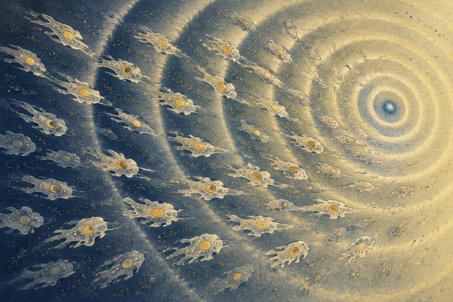
The cells do not know that food is gone in the way an animal knows hunger. Yet starvation transforms their society. Amoebae that had been crawling and feeding as independent units begin to send pulses of cyclic adenosine monophosphate, or cAMP. Nearby cells detect a pulse, produce more of the same signal and relay it onward. A chemical wave spreads through the colony. Thousands of solitary lives start to keep time together.
A new paper in Scientific Reports follows that transition in the social amoeba Dictyostelium discoideum. The eight authors are Sulimon Sattari, Md. Motaleb Hossain, Udoy S. Basak, Mikito Toda, Takeharu Nagai, Satoshi Sawai, Kazuki Horikawa and Tamiki Komatsuzaki. Their affiliations span Hokkaido, Tokushima, Tokyo and Osaka universities, with additional links to institutions in Bangladesh and Germany.
The team describes the behavior as “collective surfing.” The phrase is vivid but requires care. The cells are not passively carried in the direction of a moving wave. As a cAMP crest approaches, they actively crawl toward it—almost opposite the wave’s direction of propagation. After the crest passes, they do not turn around to chase the departing high concentration. Their speed falls, their directions scatter, and they wait for the next approaching signal.
The organism is an amoeba, not a fungus
The English name “slime mold” and the Japanese name kiiro-tamahokorikabi both invite the wrong mental picture. D. discoideum is not a fungus. It belongs to Amoebozoa and is often called a social amoeba. In well-fed conditions, haploid cells live separately in soil, consume bacteria and divide. When bacterial food disappears, the social phase begins.
Cells aggregate, form a mound and then a mobile, multicellular slug. Development culminates in a fruiting body with a stalk supporting a mass of spores. Some cells become the structure; others become the next generation. The shift from one cell to coordinated body makes Dictyostelium a compact experimental system for studying chemotaxis, signaling, cell movement, differentiation and self-organization.
Its simplicity is useful, but it does not make an amoeba a miniature human tissue. The shared value lies in questions and cellular machinery: How does a moving cell read a chemical gradient? How do local signals become a population-scale pattern? How does a crowd choose a common direction without a central controller?
One fluorescent movie, read at two spatial scales
The new work begins with a clever act of reuse. The cells expressed Flamindo2, a genetically encoded fluorescent indicator whose brightness changes with intracellular cAMP. In an unaltered frame, the mottled fluorescence preserves fine cellular structure. In a strongly blurred version of the same frame, individual cells disappear while the broad cAMP pattern remains.
The researchers applied particle image velocimetry, or PIV, to both versions. PIV was developed to recover motion in fluids: divide consecutive images into local windows, use cross-correlation to find how a particle pattern shifts, and express the result as a velocity vector field. Here, vectors from the original images represented motion at the cellular scale. Vectors from images blurred over a much larger scale represented propagation of the chemical wave.
This avoids one of the standard problems in a dense colony. Individual cell tracking requires the software to preserve a cell’s identity from frame to frame. As cells crowd and overlap visually, trajectories become fragile. PIV asks a different question: how did the local image pattern move? It can measure a field even when it cannot name every mover.
The cost is equally important. A velocity field cannot tell the complete history of a particular cell. It does not by itself reveal a leader, identify a non-responder or prove which molecular pathway caused the move. The method trades identity for continuity across a dense field.
A changing wave needs a clock that can change with it
Direction was only half the problem. The team also needed to know whether a local area was seeing the rise, crest, fall or trough of a cAMP cycle. The wave’s frequency changes as the colony develops, and the fluorescence carries smaller fluctuations and noise. A fixed-period filter would lose that evolution.
The authors therefore used wavelet analysis to assign a local phase. Unlike a conventional Fourier summary that emphasizes frequencies across a whole record, a wavelet can retain when an oscillatory component appears and how its frequency changes. That phase was then compared with the direction and magnitude of the PIV vectors.
| Layer | Operation | What it measures | What it cannot establish |
|---|---|---|---|
| Original image + PIV | Extract local displacement from fine fluorescent structure | Cell-scale velocity and direction | A continuous identity and lineage for every cell |
| Gaussian blur + PIV | Suppress cell outlines and follow broad intensity bands | Propagation of the cAMP wave | Extracellular cAMP concentration directly |
| Wavelet phase | Resolve a changing oscillation in time | Approach, crest, passage and trough | The molecular cause of each response |
The recorded field was divided into a 40-by-40 grid for the phase-resolved statistics. A representative grid element measured 134 by 162 micrometers and contained roughly 50 cells. Measurements from regions that happened to share a phase were pooled across a moving time window. That produces a strong spatial-temporal sample within the movie. It does not produce another colony.
The crest approaches; the cells row toward it
Early after starvation, before an organized cAMP wave emerged, cell movement had no clear relationship to the nominal signal phase. The relationship developed with the wave. Once the spiral pattern was mature, cells accelerated and aligned toward the positive cAMP gradient on the rising side of an approaching crest.
The wave itself propagated away from its organizing region. The cells moved toward that region. Their velocity field and the coarse-grained wave field were therefore nearly antipodal during the rising phase, approaching an angle of 180 degrees. This is the central visual result: one image scale reveals the outward-moving message; another reveals cells walking inward to meet it.
After the peak, the spatial gradient reverses. A simple “always move uphill” rule would make a cell reverse and erase its progress. It did not. The cells continued briefly in their previous direction, then became slower and less aligned in the low-gradient trough. The next approaching wave restored coordinated motion.
At the strongest coarse-graining used to isolate the wave, the Gaussian scale was about 231 micrometers, compared with a typical cell size of roughly 10 micrometers. That 231-micrometer value is a spatial smoothing scale, close to the width of the spiral band. It is not the speed of the wave. Blur beyond the useful scale eventually erases the pattern the method is trying to recover.
A long puzzle behind one short movement
Kenneth B. Raper described D. discoideum as a new species in 1935. John Tyler Bonner experimentally demonstrated a role for chemotaxis in aggregation in 1947, when the attracting substance was still known generically as acrasin. In 1967, Theo M. Konijn, Bonner and colleagues identified potent attracting activity in cAMP. Kenneth J. Tomchik and Peter N. Devreotes visualized cAMP waves across aggregating cells in 1981.
Those discoveries created a paradox. A traveling concentration wave has a front and a back. The gradient points toward the crest as it arrives and toward the same crest as it leaves—which means the direction reverses at a stationary cell. If the amoeba simply moves toward higher concentration at every moment, it should walk forward and then backward. Net aggregation should fail.
Research led by Satoshi Sawai and colleagues at the University of Tokyo supplied an important part of the answer in 2014. Cells transmitted a motility signal strongly when cAMP was increasing but not when it was falling, a rectifying behavior analogous to one-way electrical conduction. In 2024, a one-dimensional periodic-stimulation experiment showed that chemotaxis depended on the temporal gradient and that cells aligned with an incoming front without following it backward.
The 2026 paper does not replace that molecular and controlled-channel work. It changes scale. The authors follow an unconstrained two-dimensional colony from roughly two hours after starvation until about hour 17, using one analysis framework before visible coordination and after a spiral wave has developed.
1935 Raper describes D. discoideum.
1947 Bonner demonstrates chemotaxis during aggregation.
1967 Konijn, Bonner and colleagues identify cAMP’s attractant activity.
1981 Tomchik and Devreotes visualize cAMP waves.
2010 Stochastic single-cell pulses are linked to the onset of collective oscillation.
2014 Rectified directional sensing explains why cells ignore the departing wave.
2024 Periodic stimulation in a narrow channel tests temporal-gradient dependence.
2026 Multiscale PIV maps wave phase and motion across a long 2D record.
What the surfing metaphor conceals
A surfer is carried with a water wave. These cells crawl against the cAMP wave’s propagation, toward the incoming crest and its source. Their movement is metabolically active. The useful part of the analogy is timing: wait in the trough, orient when the next wave becomes legible, accelerate, then pause.
There is a second complication. Flamindo2 reports cAMP inside cells, while chemotaxis responds to cAMP outside them. Earlier measurements have disagreed about the relative timing of those signals. The paper explicitly acknowledges that what looks like cellular inertia after the intracellular crest may instead reflect an extracellular gradient that decays more slowly and is not directly measured by the reporter.
The study therefore does not prove that a cell stores the direction as an internal memory and coasts after the crest. It quantifies a relationship among intracellular fluorescence, inferred wave phase and motion. Simultaneous extracellular measurement would help distinguish persistence in the environment from persistence in the cell.
Nor does the analysis locate a new receptor or signaling molecule responsible for the behavior. It is a systems-level description that agrees with existing work on cAMP sensing and rectification. Causal perturbation—alter the wave, disable a pathway, predict the changed vector field—remains the stronger test.
Human biology is a destination, not a result
The four-university release points to wound repair, immune-cell gathering, development, cardiac behavior and other wave-governed phenomena. That relevance is plausible at the level of collective dynamics. Epithelial sheets use propagating ERK activity during coordinated migration. Human neutrophils can recruit one another through self-amplifying signal relays. Dense tissues pose the same measurement problem: local cell motion and population-scale information occupy one image at once.
But these systems do not share one universal “chemical wave.” The molecules, receptors, geometry, mechanics and biological purpose differ. The new experiment contains no human cells, wound, tumor or heart tissue. It establishes no therapy and no medical benefit.
The transferable object is initially the analytical strategy. A researcher with a suitable live reporter could blur an image across several spatial scales, recover local and collective velocity fields, assign wave phase and ask whether motion changes at a particular point in the cycle. That may help when individual tracking collapses under cell density.
Every new system would need its own calibration. A blur scale that isolates a 231-micrometer cAMP band in an amoeba colony is not a biological constant. The reporter must correspond to the relevant signal. PIV vectors must be validated against actual motion. A phase relationship must survive changes in resolution, filtering and window size.
The next colony matters more than the metaphor
The paper’s candor about its sample changes how the result should be read. One colony is not disqualifying for a method-development study, especially when the record is long and spatially rich. It is, however, a firm boundary around claims of universality. Sixteen hundred grid regions in one movie do not become 1,600 independently grown organisms.
A strong replication program would repeat the entire path: independently culture colonies, vary density and strain, image on different days, lock analysis parameters before looking at outcomes, and test whether the same phase-dependent directionality appears. It would report failed colonies and waves that never organize, not only the clean spiral.
- Biological replication: repeat the experiment in independently prepared colonies and, ideally, another laboratory.
- Condition robustness: vary cell density, strain, temperature and starvation protocol.
- Dual signaling: measure intracellular and extracellular cAMP dynamics together.
- Analytical sensitivity: publish how conclusions change with blur, grid, phase and time-window choices.
- Causal perturbation: manipulate wave timing or signaling machinery and predict the altered motion.
The underlying idea remains beautiful. Coordination does not require every cell to see a distant destination. Each cell needs only to read the local rise of a signal that the colony itself relays. The population creates a moving clock; motion is organized by phase.
That is what the single colony has shown in exceptional detail. Whether it has revealed a general rule will be decided by the less glamorous work that follows: a second colony, then a third, analyzed before anyone knows whether the picture will be as clean.
- Sattari et al. — Collective surfing of single cells on a chemo-attractant wave using multiscale Eulerian velocity vector field, Scientific Reports (2026)
- Article DOI: 10.1038/s41598-026-61774-2
- Hokkaido University — research announcement, August 26, 2026
- Hokkaido, Tokushima, Tokyo and Osaka universities — full joint release
- Tokushima University — research announcement
- University of Tokyo — research announcement
- PubMed — article record, authors, abstract and conflict statement
- Crossmark — publication history and Version of Record
- Toho University — official description of social amoebae and D. discoideum
- dictyBase — development and cAMP-guided aggregation
- American Philosophical Society — Raper’s 1935 species-description record
- Bonner — experimental evidence for chemotaxis in aggregation (1947)
- Konijn et al. — attractant activity of cAMP (1967)
- Tomchik and Devreotes — visualization of cAMP waves (1981)
- Eichinger et al. — the D. discoideum genome (2005)
- Sawai et al. — an autoregulatory circuit for long-range self-organization (2005)
- Gregor et al. — onset of collective behavior in social amoebae (2010)
- University of Tokyo and JST — official release on rectified cAMP sensing (2014)
- Nakajima et al. — Rectified directional sensing in long-range cell migration (2014)
- Odaka et al. — the Flamindo2 fluorescent cAMP indicator (2014)
- National Institute for Basic Biology — ERK waves and collective epithelial migration (2017)
- Ichimura et al. — trans-scale imaging of more than one million cells (2021)
- Ford et al. — periodic long-range signaling and morphogenesis (2023)
- Basak et al. — PIV compared with underlying collective agents (2023)
- Karmakar et al. — temporal-gradient dependence under periodic stimulation (2024)
- Strickland et al. — self-extinguishing relay waves in human neutrophil swarming (2024)
- Kakizuka et al. — mesoscale heterogeneity in spiral-wave formation (2025)
Editorial note: The organism name, paper title, author list, affiliations, Japanese institutional names, readings and technical terminology follow the original article and the joint Japanese release. No quotation has been reconstructed or translated. “Collective surfing” is attributed to the paper. The experiment comprised one colony and one imaging run; spatial regions and repeated wave cycles are not described as independent biological replication. Wound healing, immunity, development, cardiac behavior and cancer are possible application contexts for the analytical approach, not systems tested by this study. The supplied exchange-rate timestamp, August 26 at 7:24 p.m. UTC, was converted to August 27 at 4:24 a.m. Japan Standard Time.