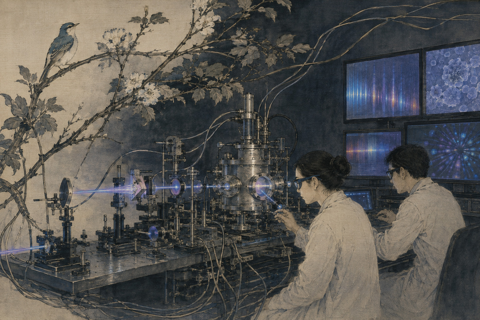
The pulse arrives once. It is over in less than one ten-trillionth of a second. By the time a conventional instrument could move a probe from one position to the next, the light is gone, the source has changed and a delicate sample may no longer be in its original state.
That is the measurement problem behind a new soft-X-ray hyperspectral microscope demonstrated at SACLA, Japan’s X-ray free-electron laser facility in Hyogo Prefecture. Instead of scanning a point across a sample, the instrument magnifies the transmitted image, slices it into 417 spatial channels and disperses every channel by photon energy onto one two-dimensional detector. One pulse produces a sparse spectral map: a spectrum associated with each sampled location.
The work was led by Yoko Takeo and Takashi Kimura of the University of Tokyo’s Institute for Solid State Physics, with Satoru Egawa of the university’s Research Center for Advanced Science and Technology, Makina Yabashi of the RIKEN SPring-8 Center and colleagues from the University of Tokyo, RIKEN and the Japan Synchrotron Radiation Research Institute. The peer-reviewed paper appeared in Optica on August 19; the University of Tokyo, RIKEN and the Japan Science and Technology Agency announced it jointly on August 20.
The achievement is easy to overstate. The prototype has not yet watched a catalyst turn over, a battery fail or a cell change chemical state. Its proof sample was a silicon-nitride film. Nor does “single shot” mean that one pulse covered every energy shown in the paper’s full absorption-edge plot. What the instrument did establish is an optical route from a femtosecond flash to spatially resolved absorption spectra—without scanning the field during that flash.
A cube of information from a flash
An ordinary monochrome microscope records intensity at horizontal and vertical coordinates. A hyperspectral microscope adds a third coordinate: wavelength or photon energy. Each location carries a spectrum rather than one brightness value. Near an X-ray absorption edge, that spectrum can be sensitive to a particular element and to aspects of its electronic or chemical state.
That extra axis matters because functional materials are rarely uniform. A catalyst can react at isolated sites. A battery electrode may age differently from particle to particle. A phase transition may nucleate in one region and spread. A biological specimen contains structures with different elemental and chemical compositions. A spectrum averaged over the whole field can erase the differences that explain how a system works—or fails.
Conventional spectromicroscopy often acquires a data cube by moving a small X-ray spot or stepping the photon energy and collecting many frames. It can be extraordinarily powerful when the object stays put. It becomes a poor match for a state that lasts femtoseconds, for a sample altered by the first exposure, or for an XFEL pulse whose spectrum is not identical to the next one.
SACLA’s soft-X-ray beam at BL1 is generated by self-amplified spontaneous emission. That makes very intense, very short light available, but individual pulses fluctuate. If an image is assembled from repeated shots, a researcher must separate true changes in the sample from changes in the illumination. The new design attacks both problems at once: record many places simultaneously, and diagnose the illumination with the same spatially resolved spectral logic.
Two mirrors preserve the image; 417 gratings spread its energies
The microscope begins with grazing-incidence Wolter mirrors. Soft X-rays do not pass through and bend in ordinary glass optics as visible light does. At shallow angles, however, they can reflect. A condenser Wolter mirror illuminates the sample, and an objective Wolter mirror forms a magnified transmitted image.
Reflection provides a decisive advantage for spectroscopy: the mirror system is achromatic in principle. A diffractive X-ray optic such as a zone plate changes focal length with wavelength. The Wolter system can preserve focus and magnification across an energy range, allowing the spectral content of the image to reach the next stage without wavelength-dependent blur or scale changes of the same kind.
That next stage is the newly fabricated multi-aperture grating, or MAG. On a 14-millimeter-square chip, the team integrated 417 apertures with corresponding transmission gratings. Each opening selects one part of the magnified image. Each grating sends different photon energies to different horizontal positions while focusing its first-order diffracted light onto the detector. The patterns are arranged so the miniature spectra remain spatially separated.
The openings measure 300 by 80 micrometers on the MAG and sit on a 600-by-400-micrometer pitch. With the microscope’s roughly 250-fold magnification, that pitch corresponds to 2.4 by 1.6 micrometers at the specimen. The grating grooves have a 300-nanometer period. Electron-beam lithography and other semiconductor processes formed the structures in silicon and silicon-nitride films at the University of Tokyo’s Takeda Super Clean Room.
From specimen to detector, the apparatus extends about 6.5 meters. This is a beamline instrument, not a benchtop microscope nearing hospital or factory deployment. The project’s immediate significance lies in experimental capability at large X-ray facilities.
| Component | Job | Measured performance or constraint |
|---|---|---|
| SACLA BL1 | Supplies a bright soft-X-ray pulse shorter than 100 femtoseconds. | Freezes one moment, but pulse spectra vary from shot to shot and across the beam. |
| Condenser Wolter mirror | Illuminates the specimen without a refractive lens. | Illumination measured 28 × 37 μm FWHM in the experiment. |
| Objective Wolter mirror | Magnifies the transmitted image achromatically. | The hyperspectral setup produced a 39 × 39 μm field. |
| Multi-aperture grating | Samples the image at 417 locations and disperses each sample. | 2.4 × 1.6 μm sampling; it is not a continuous spectrum at every camera pixel. |
| 2D detector | Records all separated spectra at once. | Grating-only resolving power was calculated as E/ΔE ≈ 1,000 near 100 eV; detector pixels further limit effective resolution. |
The light source became the first subject
Before putting a sample in the beam, the researchers used the MAG to measure the XFEL illumination itself. Three representative pulses looked different from one another, and a 100-shot average hid variations visible in the individual frames. The spectrum was not spatially uniform within a pulse, either. After averaging by spatial channel, photon energy was slightly higher near the optical axis than farther off axis.
Those results make the apparatus useful beyond microscopy. A detector that integrates the whole beam can report a pulse-average spectrum while missing local energy shifts. Spatially resolved, shot-by-shot spectra can show the light that actually struck different parts of a specimen. That helps researchers tell an illumination artifact from a material response and could aid XFEL tuning and diagnostics.
“Could” is the operative word. The paper demonstrates the principle; it does not say the device has become SACLA’s routine production monitor. Calibration speed, long-term stability, automated reconstruction, photon throughput and performance at other energies still determine whether a prototype becomes an everyday beamline tool.
The silicon-nitride edge—and a careful reading of the plot
For the material test, the edge of a silicon-nitride film covered only the lower portion of the field. That geometry put sample and reference regions in one image. Near the film’s absorption edge, the transmitted spectrum through silicon nitride could be compared with light passing through the open region during the same pulse.
At source settings of 104 and 105 eV, the light behind the film showed a lower central energy and a broader spectrum. The team attributed both changes to stronger absorption around the edge. To correct for the illumination’s spatial and shot-to-shot structure, the sample-free upper region served as a reference from which the incident spectrum at the film position was estimated. A test on sample-free data put the root-mean-square accuracy of the estimated central photon energy at 0.02 eV.
The most important editorial caveat sits in the acquisition sequence. The spatial spectrum at a given setting came from a single pulse. But the researchers repeated measurements while stepping the central energy from 97 to 110 eV in 1 eV increments to build the broad transmission curve. In other words, a pulse contains a useful energy band, not the entire plotted scan.
That distinction does not negate the result. At a chosen absorption edge, a sufficiently broad pulse can carry enough spectral structure to distinguish regions in one exposure. But it defines the next engineering target: wider usable bandwidth and greater detector and grating performance if researchers want a fuller absorption signature without stepping the source.
- Demonstrated: 417 spatially corresponding spectra recorded from one soft-X-ray pulse.
- Demonstrated: spatial and shot-to-shot variation in the SACLA BL1 beam.
- Demonstrated: a spatial transmission change in a silicon-nitride film near its absorption edge.
- Not yet demonstrated: a changing catalyst, operating battery or living cell measured for chemical state with this instrument.
- Not claimed: continuous video, three-dimensional tomography, atomic resolution or a compact commercial product.
An astronomy mirror moves into the microscope
X-ray imaging began with Wilhelm Röntgen’s 1895 discovery, but focusing the rays proved unlike building an optical camera. X-rays tend to penetrate or be absorbed rather than reflect from a mirror facing them directly. German physicist Hans Wolter showed in 1952 how successive reflections at grazing incidence could form an X-ray image. Variants of the geometry became standard in X-ray astronomy.
The University of Tokyo group has spent years adapting precise cylindrical Wolter mirrors to microscopy. In 2022, the researchers combined a Wolter mirror with ptychographic reconstruction at SPring-8 and reported roughly 50-nanometer resolution over a broad soft-X-ray energy range. That number should not be transferred to the 2026 hyperspectral prototype. The present MAG system gives up that much finer spatial detail in exchange for simultaneous spectral sampling; its reported sampling is micrometer-scale.
In 2024, members of the same collaboration paired Wolter optics with a femtosecond SACLA pulse to image living CHO K-1 mammalian cells in the soft-X-ray “water window.” A single exposure caught the cell before radiation damage developed during a later 0.5-second multi-shot exposure. That experiment supplied a frozen image. The 2026 design adds a way to distribute the image into many spectra.
1895 — Wilhelm Röntgen discovers X-rays.
1952 — Hans Wolter formalizes two-reflection grazing-incidence X-ray optics.
March 2012 — SACLA begins user operation.
July 2016 — The upgraded soft-X-ray FEL at BL1 begins user operation.
2022 — The Tokyo group reports achromatic, roughly 50 nm soft-X-ray imaging with Wolter optics and ptychography.
2024 — The collaboration reports a single-pulse soft-X-ray image of living mammalian cells at SACLA.
August 2026 — The 417-channel single-shot hyperspectral method appears in Optica.
Femtoseconds do not automatically make a movie
The instrument’s time resolution is set by the XFEL pulse duration. That means one exposure can be much shorter than motion or damage that follows. It does not mean the camera continuously records trillions of frames per second.
To reconstruct a reaction over time, researchers would generally trigger it with a pump pulse and probe it at different delays, or otherwise repeat the experiment at defined time points. The specimen must return to the same starting condition, be replaced after each shot, or provide many equivalent targets. Timing jitter, pump uniformity and the ability to align different shots then become part of the experiment.
Single-shot spatial spectroscopy still changes the balance. Every sampled location in one frame shares the same pulse history. Local differences are no longer separated by a mechanical scan, and a specimen altered by exposure has already yielded all 417 channels before that later damage can contaminate the measurement window.
The price is explicit. The MAG samples a grid rather than delivering an uninterrupted hyperspectral camera image. Its grating-limited resolving power of about 1,000 near 100 eV is a theoretical calculation for the grating—equivalent to roughly 0.1 eV before other limits—not a blanket measured specification for the complete instrument. Detector-pixel size lowers the effective spectral resolution, while the division of light among many channels puts pressure on efficiency and signal.
The authors propose broader source bandwidth, faster detectors, improved spectroscopic elements and higher repetition rates. They also discuss extensions to hard X-rays with other mirror optics and to pump-probe experiments. These are research directions, not features already delivered by the apparatus described in the paper.
A coordinate system for a future chemical map
The long-term target is compelling: identify not only where matter is, but where a specific chemical or electronic state appears during a fleeting event. In an electrode, that could distinguish regions changing valence at different rates. In a catalyst, it could connect a transient state to one microscopic zone. In biology, it could eventually map chemical differences before radiation damage rearranges the specimen.
Reaching those applications requires evidence beyond a thin-film edge. Real materials scatter, absorb and evolve in more complicated ways. Biological specimens introduce water, thickness and dose constraints. A credible chemical-state movie would need validated spectral calibration, spatial resolution, incident-beam correction, temporal synchronization and a reconstruction method whose uncertainties remain under control.
For now, the achievement is narrower and cleaner. Achromatic mirrors preserved the magnified soft-X-ray image. Four hundred seventeen integrated spectroscopic elements kept location attached to energy. The detector captured the pattern during one pulse. And the same architecture exposed variations in the XFEL that might otherwise masquerade as sample behavior.
This is not a microscope that sees everything in one shot. It is an instrument that refuses to throw away where a spectrum came from. In ultrafast science, that surviving link between place and energy may be the difference between a bright flash and a usable map.
- Japan Science and Technology Agency — joint release, “Femtosecond soft-X-ray hyperspectral microscope realized,” August 20, 2026 (Japanese)
- JST — detailed joint-release PDF with apparatus and test figures (Japanese)
- Takeo et al., “Femtosecond hyperspectral imaging with a single-shot X-ray pulse,” Optica 13, 1643–1648 (2026)
- KAKEN — research record for “Demonstration of X-ray single-shot microscopic spectroscopic imaging,” project 20H04451
- University of Tokyo Institute for Solid State Physics — 2022 Wolter-mirror and ptychography release (Japanese)
- University of Tokyo RCAST — 2024 living-cell single-pulse soft-X-ray imaging release (Japanese)
- RIKEN — SACLA Beamline Research and Development Group
- NASA Goddard — X-ray telescope optics and Hans Wolter’s 1952 designs
Attribution note: Specifications, experimental results and proposed applications come from the paper and joint release. E/ΔE ≈ 1,000 is the calculated grating-limited resolving power, not the measured end-to-end instrument resolution. The explanation that the 97–110 eV curve combines multiple source settings follows the paper’s method. The assessment of the instrument as a trade among time, space and energy is Japan.co.jp analysis.