When a mouse wakes, a researcher cannot ask, “Do you remember the room from two days ago?” The answer must be read in motion: where the animal pauses, which place it avoids and which turn it chooses.
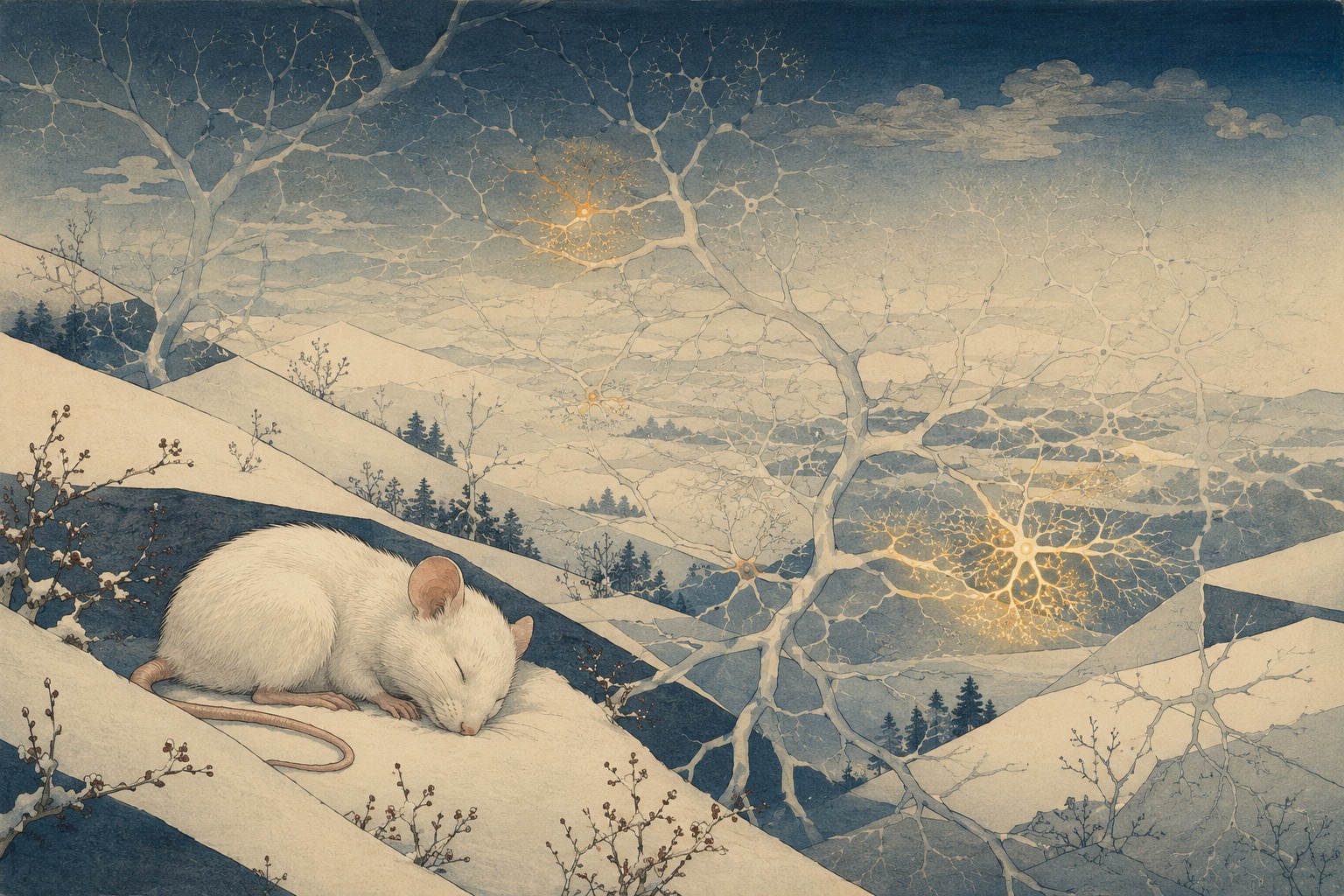
In this experiment, the answer was strangely calm. While the mouse was in an induced state of low temperature and metabolism, firing in the hippocampus fell by roughly 70%. More than half its observed synapses—the junctions through which neurons communicate—were eliminated. Once normal physiology returned, however, the animal still expressed a contextual fear memory learned before hibernation and still made learned spatial choices. In some behavioral tests, performance did not merely survive; it improved.
The result, reported in Science in August 2026 by a team centered on the Memory Research Unit at the Okinawa Institute of Science and Technology, asked a deeper question than whether a hibernating animal forgets. When a brain’s wiring is extensively remodeled, what must remain for an old memory to become accessible again? Artificial hibernation was not the destination of the research. It was the stress test.
A switch for winter in the hypothalamus
This artificial hibernation was not anesthesia, ordinary sleep or a mouse placed in a cold room. It began with a neural circuit described in Nature in 2020 by Takeshi Sakurai and colleagues at the University of Tsukuba. Activating a group of QRFP-expressing neurons in the preoptic hypothalamus drove a sustained fall in body temperature, oxygen consumption, heart rate and movement. The state became known as Q-neuron-induced hypometabolism and hypothermia, or QIH.
The investigators used chemogenetics: a designer receptor was placed selectively in Q neurons, then activated with a matching compound. The striking observation was that a hibernation-like state could also be induced in rats, which do not naturally hibernate. That suggested that something resembling a torpor program might be accessible through conserved mammalian circuitry, rather than existing only in seasonal hibernators.
QIH is not identical to the months-long natural hibernation of a ground squirrel or bear, nor is it the same as anesthesia or coma. It is a controlled, reversible hypothermic and hypometabolic state. The 2026 team used it as a perturbation powerful enough to prune a large share of connections, making it possible to ask which structures disappear, which survive and which can be rebuilt after normal physiology returns.
Three ways to ask a mouse what it remembers
Memory is not a single measurement. The researchers aligned evidence at three scales—behavior, population activity and synaptic ultrastructure. Each scale answered a different question and imposed a different limit.
| Scale | What was measured | What it showed—and what it could not |
|---|---|---|
| Behavior | Contextual fear conditioning and spatial choice: whether a mouse froze in a learned context and chose a learned location | Access to the learned information survived QIH. These tasks do not reproduce human autobiographical memory |
| Population | Hippocampal firing, place-cell activity and spatial representation | Overall activity plunged, but the representation of place was preserved after QIH. This was not a reading of every memory across the brain |
| Connection | Two-photon imaging, fluorescent labeling and electron microscopy of dendritic spines and engram-to-engram synapses | Many connections vanished while clustered engram synapses were selectively spared. The whole sequence was not filmed across every synapse of one living brain |
In contextual fear conditioning, a mouse learns an association between a particular environment and an aversive event. When it later returns and becomes still—a response known as freezing—that behavior serves as evidence that the context was remembered. A separate spatial task tested whether it could repeat a learned location or choice. A hippocampal-lesion experiment helped establish that recall after QIH still depended on the hippocampus rather than being rerouted entirely around it.
The team also followed place cells, hippocampal neurons that fire strongly when an animal occupies a particular location. Together they form an internal map. Activity was profoundly suppressed during the induced state, but after recovery the learned spatial representation returned. That makes the case stronger than a coincidental repetition of behavior, while still falling short of revealing the subjective content of the memory.
The 120-year search for a physical memory
The idea that experience must leave a physical trace is more than a century old. In 1904, German biologist Richard Semon named that trace the “engram.” But the proposed object could not be seen. Was a memory held by one cell, one brain region, one synapse, or an arrangement distributed across all three?
1904 Richard Semon calls the physical trace left by experience an “engram.”
1920s–1950s Karl Lashley lesions different parts of the cortex in maze-trained rats, fails to find one memory center and argues for distributed storage.
1949 Donald Hebb proposes cell assemblies and the rule that connections strengthen when neurons are repeatedly active together.
1957 Scoville and Milner report patient H.M., establishing the medial temporal lobe’s essential role in forming new long-term memories.
1973 Bliss and Lømo describe long-term potentiation, a durable increase in synaptic transmission in hippocampal pathways.
2012 Susumu Tonegawa’s group optogenetically reactivates hippocampal cells tagged during learning and triggers fear-memory recall.
2025 Three-dimensional electron microscopy reconstructs synaptic architecture within a mouse hippocampal engram.
2026 The OIST-led study strips away large numbers of connections with QIH and compares the surviving architecture with retained memory.
Lashley’s famous “search for the engram” looked like a failure, but it left an enduring clue: a memory did not behave like a document filed in a single cortical drawer. Hebb supplied a mechanism of assembly—neurons that repeatedly act together become more strongly linked. The case of H.M. established the hippocampal system’s importance in human memory, and long-term potentiation showed how activity could leave communication at a synapse durably altered.
In 2012, researchers could finally tag hippocampal neurons active during learning and later reactivate them with light. The animal expressed the associated fear response. The engram had moved from a theoretical trace to a cell population that could be labeled and manipulated. Yet another question remained: which connections among those cells preserve a memory over time?
Why “the biggest spine wins” was not enough
The history of long-term potentiation makes a simple intuition attractive: a stronger synapse, or a larger dendritic spine, should be the durable one. Dendritic spines are tiny protrusions on the receiving branches of a neuron, and many excitatory synapses form on them. Larger spines often have larger postsynaptic structures and tend, on average, to support stronger transmission.
QIH did not spare only the large spines. Large and small structures were both removed, and their size before the induced state did not reliably predict survival. Yet the memories endured. That does not mean synaptic strength is irrelevant to learning. Potentiation can remain crucial when an experience is first encoded. The new result instead separates two problems: the mechanism that writes a memory and the architecture that allows an established memory to survive long periods of change.
- Encoding: turning an experience into a new pattern of activity and connection change. Long-term potentiation may be central here.
- Retention: preserving enough information to reconstruct the memory across time and turnover. This is the 2026 paper’s central question.
- Retrieval: reactivating that information from a cue and expressing it in behavior. A retained memory can remain hidden if retrieval fails.
What survived was not one line, but an arrangement
The team isolated connections linking neurons that had been active during learning—the engram cells—and examined their fine structure. The key technique was correlative light and electron microscopy, or CLEM. Fluorescence identified which cells and contacts belonged to the sparse engram population. Electron microscopy then resolved membranes, spines, postsynaptic densities and terminals at nanometer scale. Aligning the two maps let researchers recognize an engram-to-engram synapse inside a vast field of ordinary connections.
The connections most likely to survive were not isolated. They were engram synapses grouped close together along a dendrite. Contacts associated with multi-synaptic boutons, or MSBs, were also preferentially conserved. An MSB is a single presynaptic terminal that contacts more than one dendritic spine, sometimes across more than one receiving cell. It resembles a junction feeding several nearby routes, rather than a lone bridge carrying all the traffic.
A 2025 Science study had reconstructed the three-dimensional synaptic architecture of hippocampal engrams and described multiple contacts through MSBs alongside highly local structural changes after learning. The 2026 study viewed that architecture from a different angle—not as a structure formed during learning, but as one selectively left standing after widespread synaptic loss. Memory’s durability may lie less in the toughness of any single part than in higher-order wiring through which parts reinforce one another.
Did a vanished connection truly disappear?
Live two-photon imaging showed many dendritic spines disappearing during QIH and new connections forming after the animal returned to normal. The study’s public data and preprint also describe lost spines reappearing at or near previous locations. The brain may not need to preserve every original component if surviving architecture can serve as a scaffold for rebuilding a similar circuit.
There is an important technical limit. Electron microscopy requires fixed, sectioned tissue. It cannot follow the same living synapse before, during and after QIH. The behavioral tests, live spine imaging, neural recordings and CLEM therefore integrate carefully designed cohorts and methods. Researchers did not watch every connection disappear inside one transparent brain and then test the same tissue after recall.
That does not erase the result. Its strength is convergence: behavior, spatial representation, synapse loss and selective structural preservation point in the same direction. Its remaining weakness is causality. If the clustered architecture is selectively disrupted, does the memory alone fail? If a cluster is artificially built, does retention improve? Those interventions would turn an association into a mechanism.
Natural hibernation refuses a simple answer
Biologists have long known that hibernating brains remodel. Dendrites retract, synaptic proteins change and structures can return during arousal. But memory findings across hibernators are mixed. Depending on species, duration, task and testing time, researchers have observed impairment, preservation and, after brief torpor, even enhancement on particular learning measures.
That variation is one reason QIH is valuable. Natural hibernation entangles season, nutrition, age and ecology. An induced state allows timing, duration, learning task and control groups to be held more tightly. The 2026 work included animals that did not undergo QIH and anesthesia controls, helping distinguish the induced metabolic state from the mere fact of remaining inactive.
But a roughly 48-hour laboratory state cannot simply be extrapolated to months of natural hibernation or to clinical hypothermia. Resilience differs across brain regions. Young, healthy laboratory mice are not older humans, patients with neurodegenerative disease or people recovering from traumatic injury.
So where was the memory?
The most careful answer is: not in one place. A memory spans hippocampal engram cells, selected connections among them, the local arrangement of those contacts, population-level spatial activity and, over time, representations distributed into cortex. The artificial-hibernation experiment exposed one level of that hierarchy that resisted massive loss in the total number of contacts.
At least four explanations remain plausible. A sparse set of clusters may directly preserve the essential trace. The clusters may act as seeds from which a circuit is regrown. Redundancy in a distributed network may compensate for missing connections. Or cortical information may provide a cue that helps reconstruct hippocampal patterns. These possibilities can coexist.
Yu-Ju Lin, the paper’s first author, emphasized in OIST’s account that the structural pattern was correlated with retention; a causal link has not yet been established. That distinction is not a footnote. “Associated with” is the scientifically important phrase. “Stored the memory” would reach beyond the evidence.
What the study does not mean
- It does not show that humans can be placed safely into artificial hibernation. A clinically usable counterpart to Q-neuron manipulation has not been established.
- It is not a treatment that restores memories lost to Alzheimer’s disease or injury. Neuronal death, pathological proteins and inflammation are different from reversible QIH remodeling.
- It does not show that half the brain’s synapses are disposable. It shows that total number and behavioral performance did not move in simple proportion under these experimental conditions.
- It does not overturn long-term potentiation. Encoding a memory and retaining an established trace are different scientific questions.
- Fear conditioning and spatial choice are not tests of a human childhood, language or identity.
- Finding a cluster is not a technique for reading, transplanting or digitally backing up a memory.
Artificial hibernation has inspired proposals to lower oxygen demand after severe injury, protect organs or support long-duration spaceflight. The 2020 QIH discovery raised those possibilities as future directions. If such a state could ever be controlled safely in humans, the stability of memory circuits during hypometabolism would be a central safety question. For now, this remains basic research based on genetic manipulation of particular circuits in mice and rats.
How a changing brain keeps the same past
The broad problem behind OIST’s Memory Research Unit is memory in a changing brain. Synapses appear and disappear every day. Their proteins turn over. The activity of a neuronal population drifts. Yet a route learned yesterday remains walkable today. Stability emerging from unstable material is one of neuroscience’s deepest puzzles.
Artificial hibernation accelerated ordinary change into a severe test. Removing many connections did not reduce the memory to zero. That invites a shift in metaphor—from the brain as an indestructible recording medium to the brain as an organization able to rebuild.
Imagine an old road to a temple. Pavement breaks, several bridges fall and signs disappear. If the arrangement of the forks, the relationship between valley and ridge, and several neighboring paths remain, a traveler can still find the destination. A memory may resemble that terrain more than an inscription on a stone.
OIST’s mice revealed a structure that survived change, not an immutable component. The next step is to disrupt, create and follow that structure over longer periods. If those experiments establish causality, the science of long-term memory will have moved from counting how many connections remain to explaining how the surviving connections are organized.
Sources and reporting basis
- OIST, “Artificial hibernation reveals secrets of long-term memory” (August 13, 2026)
- Lin et al., “Artificial hibernation reveals synaptic engram architecture associated with memory retention,” Science (2026)
- Dryad source data, analysis scripts and experimental variables for the study (published June 30, 2026)
- The study’s bioRxiv preprint (2025)
- Takahashi et al., “A discrete neuronal circuit induces a hibernation-like state in rodents,” Nature (2020)
- University of Tsukuba, explanation of Q-neuron-induced hypometabolism and hypothermia (June 12, 2020)
- OIST Memory Research Unit, research program and methods
- Josselyn, Köhler and Frankland, “Heroes of the Engram,” Journal of Neuroscience (history of the engram, 2015)
- Scoville and Milner, “Loss of recent memory after bilateral hippocampal lesions” (patient H.M., 1957)
- Bliss and Lømo, “Long-lasting potentiation of synaptic transmission…” (long-term potentiation, 1973)
- Liu et al., “Optogenetic stimulation of a hippocampal engram activates fear memory recall,” Nature (2012)
- Josselyn and Tonegawa, “Memory engrams: Recalling the past and imagining the future,” Science (2020)
- Choi et al., “Synaptic architecture of a memory engram in the mouse hippocampus,” Science (2025)
- U.S. National Institutes of Health, account of the 2025 hippocampal engram-architecture study
- von der Ohe et al., “Synaptic Protein Dynamics in Hibernation,” Journal of Neuroscience (reversible hibernation remodeling, 2007)
- Horowitz et al., “Torpor enhances synaptic strength and restores memory performance…” Scientific Reports (2021)
Editor’s note: “More than half” refers to synapses observed in the mouse hippocampus under the paper’s experimental conditions, not the whole brain and not humans. The public preprint and data set were used to clarify methods; the peer-reviewed paper and OIST’s official account govern the reported conclusions. This article is based on material available by 3:14 a.m. Japan Standard Time on August 15, 2026.