- A cell is not a still photograph
- The numbers behind the advance
- Chemistry without fluorescent labels
- Why adding depth changes the biology
- Eleven angles, one fast volume
- A second experiment: scanning chemical signatures
- The long route from infrared images to moving volumes
- The people and institutions behind the instrument
- Lipid droplets are an important first test
- What the images do not yet tell us
- A window into the cell’s moving chemistry
A cell is not a still photograph
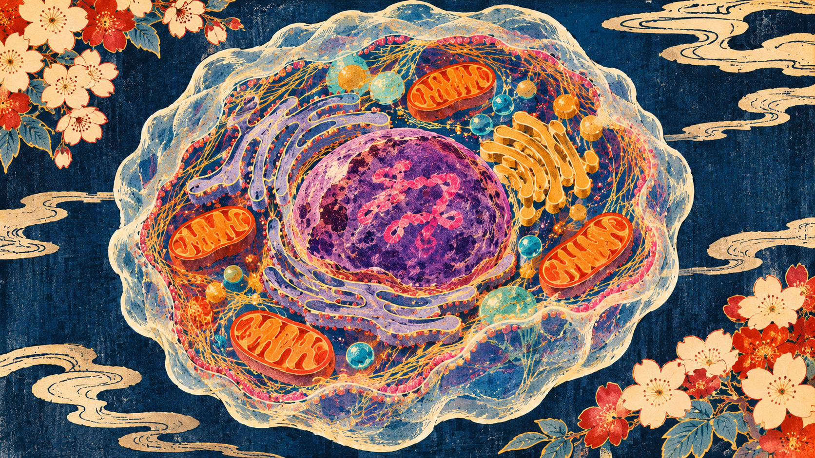
A living cell is a crowded, moving three-dimensional environment. Lipid droplets travel, structures assemble and dissolve, and chemical composition varies from one region to the next. A static microscope image can freeze a moment; a conventional two-dimensional movie can follow motion but may lose whatever happens along the depth axis. Fluorescent markers reveal selected components, yet labeling is not always desirable or sufficient. A team at the University of Tokyo and RIKEN has reported a way to narrow those compromises.
Announced November 2 and described in a peer-reviewed Nature Communications paper, the instrument combines mid-infrared photothermal measurements with optical diffraction tomography. It reconstructed 19.2 chemically contrasted three-dimensional volumes every second, allowing researchers to follow lipid droplets inside living cells without fluorescent staining. This is not a camera looking directly into a cell. It is a carefully synchronized optical measurement and reconstruction system.
The numbers behind the advance
At 19.2 volumes per second, a complete volumetric acquisition takes roughly 52 milliseconds. The team reports a nearly 400-fold improvement over previous implementations of three-dimensional mid-infrared photothermal tomography, which had remained below about 0.05 volumes per second. It measured lateral resolution near 0.35 micrometers and axial resolution near 1.2 micrometers in its live-cell experiments. The paper also reports signal-to-noise ratio exceeding 70 without temporal averaging.
Those figures are meaningful only with the correct comparison. A volume is one reconstructed three-dimensional dataset, not one photograph or a full chemical identification of everything in the cell. The reported throughput concerns the configured experiment. It should not be extrapolated uncritically to thick tissue, a whole organ, or a clinical scanner.
Chemistry without fluorescent labels
Molecules absorb mid-infrared light at frequencies related to their chemical bonds. Lipids, for example, carry characteristic C–H vibrations. Choosing a corresponding infrared wavenumber offers chemical contrast: structures enriched in those bonds respond differently from their surroundings. But directly imaging long-wavelength infrared light entails a resolution penalty.
Mid-infrared photothermal microscopy finds a way around part of that difficulty. Infrared absorption slightly heats the sample and changes its refractive index. Visible light probes that minute optical change. The technique therefore uses infrared absorption to select a chemical signature and visible-light optics to measure its spatial distribution. This is chemically informative, not a universal molecular barcode. Overlapping spectra and complex biological samples still require careful interpretation.
Why adding depth changes the biology
Consider a droplet disappearing from a microscope image. It may have moved sideways, moved toward or away from the objective, merged with another object, or changed in ways that alter its contrast. A two-dimensional trajectory cannot always distinguish these possibilities. The new method provides a three-dimensional track and makes motion along the optical axis observable.
The investigators recorded lipid droplets within living cells for approximately 3.6 seconds. Their trajectories showed features of anomalous, specifically more constrained, diffusion rather than the spreading expected from ordinary free diffusion. The behavior also varied across intracellular locations. That is a measurement of motion, not by itself an identification of every force or biological mechanism producing the motion.
Eleven angles, one fast volume
The researchers used intense nanosecond infrared pulses to obtain photothermal contrast with adequate signal from a single exposure at each angle. A spatial light modulator changed the visible illumination direction roughly every 2.3 milliseconds. Eleven illumination angles contributed information for each reconstructed volume, with the laser, camera and illumination apparatus precisely synchronized.
The system compared measurements taken with infrared excitation on and off, then recovered the three-dimensional distribution of photothermal response through optical diffraction tomography. Reconstructing a volume from multiple optical measurements is the essential distinction from conventional photography. Improved sensitivity and rapid angular scanning jointly enabled the much faster result.
A second experiment: scanning chemical signatures
The team also varied the infrared wavenumber across an approximately 300 cm⁻¹ spectral window, collecting roughly 20 spectral sampling points worth of three-dimensional images in about a second. On fixed cells, different regions—including lipid droplets and nucleoli—displayed distinct vibrational spectral responses. A nucleolus is a nuclear structure involved in making the machinery of protein synthesis.
This was a separate demonstration from continuous live-cell tracking at a selected lipid-sensitive wavenumber. The paper does not establish that a microscope can simultaneously identify every chemical species throughout a living cell at 19.2 complete spectra per second. Distinguishing those claims matters because chemical specificity, imaging speed and biological applicability place different demands on the equipment.
The long route from infrared images to moving volumes
The new achievement is part of a line of work rather than a sudden leap from nowhere. In July 2023, University of Tokyo researchers reported high-speed, label-free single-cell mid-infrared microscopy capable of watching molecular dynamics. In April 2024, another university announcement described mid-infrared microscopy with approximately 100-nanometer spatial resolution for visualizing molecular vibrational distributions.
Those projects emphasized different performance dimensions and should not be ranked as though they were identical devices in a race. Together they illustrate the challenges of chemical selectivity, spatial detail and temporal speed. The 2026 work advances the ability to preserve useful chemical contrast while acquiring depth-resolved information quickly enough to track motion.
The people and institutions behind the instrument
The published authors are Masato Fukushima, Keiichiro Toda, Yusei Sugawara, Shotaro Kawano and Takuro Ideguchi. The University of Tokyo identifies Fukushima and Sugawara as graduate researchers, Toda as a project assistant professor, Kawano as a project researcher and Ideguchi as a professor in its Graduate School of Frontier Sciences who also directs a research team at the RIKEN Center for Advanced Photonics.
It is a collaboration across physics, photonics, measurement and computational imaging. Its contribution is not simply a faster electronic shutter: the optical signal, illumination sequence, computational reconstruction and biological validation had to work together. The accompanying research paper gives readers a way to assess those choices rather than relying on a promotional description of the technology.
Lipid droplets are an important first test
Lipid droplets store neutral lipids and participate in cellular energy management and interactions with other organelles. Their chemistry is favorable for mid-infrared contrast, and their movements make them useful test objects for evaluating three-dimensional tracking. Measuring them inside living cells is therefore a meaningful proof of capability, even if it is not a comprehensive survey of cell biology.
Possible future questions include how intracellular cargo is transported, how lipid-rich structures form or merge, and whether changes in molecular organization accompany the formation of biomolecular condensates. These are directions for subsequent experiments. The study did not report a validated disease diagnosis, new drug, or proven clinical benefit.
What the images do not yet tell us
More detail is not automatically more understanding. A trajectory may reveal that movement is constrained, but determining why could require genetic interventions, orthogonal microscopy or biochemical assays. Similarly, differences between local infrared spectra do not necessarily identify a unique molecule amid a complex mixture.
Practical limitations also matter. The experiments rely on specialized infrared sources, rapid wavefront modulation, synchronized cameras and computational reconstruction. Questions remain about usable sample thickness, wavelength coverage, sustained imaging, illumination-related biological effects, cost, throughput and reproducibility in other laboratories. The paper is a substantial technical result, not evidence that this configuration is ready for routine clinical deployment.
A window into the cell’s moving chemistry
The achievement is best understood as a new kind of measurement rather than an immediate medical product. Instead of deciding between a sharp chemical snapshot and a less informative movie, researchers have demonstrated a way to collect rapidly changing three-dimensional maps of a selected molecular signature.
If the method becomes more accessible, it could help investigators ask increasingly precise questions about what substances are present, where they move and how their motion changes. For now, its achievement is already clear and narrower: a peer-reviewed experiment captured live-cell lipid-droplet motion in three dimensions at video-rate volumetric speed, with chemically meaningful contrast and no fluorescent label.
Sources and reporting method
This documentary report compares the peer-reviewed original paper with the institutions’ published disclosures and earlier research announcements.
- University of Tokyo Graduate School of Frontier Sciences, Nov. 2, 2026
- RIKEN, joint research announcement
- Nature Communications, peer-reviewed research paper
- University of Tokyo, 2023 related microscopy research
- University of Tokyo, 2024 related microscopy research
- University of Tokyo Department of Physics, English research announcement