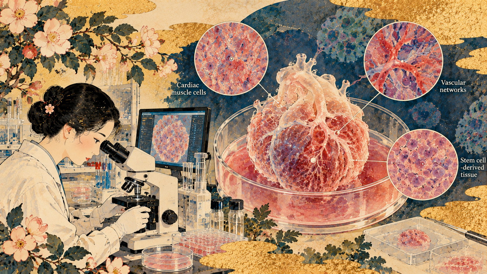
The hidden change between day 28 and day 60
A piece of human heart tissue grown in a laboratory can beat. But what happens if scientists keep it alive long enough for an inherited disease to reveal itself? In engineered tissue made from cells of a patient with Hutchinson–Gilford progeria syndrome, an abnormal protein undetectable at day 28 was present by day 60. The tissue also became less likely to respond to electrical pacing. Researchers at Kyoto University’s Center for iPS Cell Research and Application, working with international collaborators, have turned an elusive feature of a rare disorder into something they can watch develop. It is an advance in modeling disease, not evidence of a cure.
A paper in August, a university announcement in October
The peer-reviewed paper appeared online in Biofabrication on August 28, 2026. Kyoto University’s CiRA research announcement followed on October 26, and the university listed it among research news on October 27. Those dates matter: the new October reporting concerns publication and explanation of an earlier scientific paper, not a trial completed this week. The paper is titled “Engineering epicardium-integrated human iPSC-derived heart tissue for modelling Hutchinson-Gilford progeria syndrome.”
Why a premature-aging disorder attacks the cardiovascular system
Hutchinson–Gilford progeria syndrome, or HGPS, is a rare genetic disorder most often associated with an alteration in LMNA. It leads to production of progerin, an abnormal protein affecting the nuclear envelope and cellular function. Children develop features resembling accelerated aging, and severe cardiovascular complications are central to the disease. The disorder is not simply ordinary aging happening faster: the mutation produces a specific, destructive molecular process. Reconstructing the heart’s involvement has been particularly difficult.
Twenty years after a Japanese stem-cell milestone
In 2006, Shinya Yamanaka and colleagues showed that mouse adult cells could be reprogrammed into induced pluripotent stem cells. Human iPS cells followed in 2007. The achievement opened two broad routes: producing specialized cells for possible therapeutic use, and growing patient-derived cells in the laboratory to investigate disease or evaluate drug candidates. For rare disorders, the second route can be especially valuable because obtaining living, diseased human heart tissue is extraordinarily difficult. The new Kyoto study builds on that historical turn in biomedical science.
Why heart muscle alone is not enough
Heart muscle cells generate contraction, but they are only part of the organ. Fibroblasts help maintain its supporting structure, smooth-muscle cells contribute to vascular function, and interactions among these and other cell types influence disease. Conventional flat cultures can isolate a mechanism, but often miss the complexity of neighboring cells and the physical environment. The researchers therefore added epicardial cells—descendants of the heart’s outer layer that can produce other cardiac support-cell types—to their engineered tissue.
Building a three-dimensional heart model
Using induced pluripotent stem cells derived from a patient with HGPS and the patient’s healthy parents, the team generated cardiac muscle cells and epicardial cells. They combined them in an extracellular-matrix gel to make contracting engineered heart tissue, known as EHT. Within this system, epicardial cells differentiated into cardiac fibroblasts and vascular smooth muscle cells. The resulting structure had a more compact organization than tissue containing cardiomyocytes alone. Crucially, it allowed researchers to follow a disease-related process across two months in a multicellular human system.
What adding epicardial cells changed
The university reports contraction stress of 46 ± 8 micronewtons per square millimeter in cardiomyocyte-only constructs (six tissues), compared with 138 ± 90 in epicardium-integrated constructs (27 tissues). These are measurements in engineered tissue, not clinical assessments of a person’s heart. They nevertheless demonstrate why the cellular composition of a laboratory model matters. A more realistic array of interacting cells may reveal changes that simpler preparations do not capture.
Progerin arrives late
In the patient-derived constructs, progerin accumulation was not detectable at day 28 but was found by day 60. Researchers also observed changes in gene expression associated with oxidative stress, atherosclerosis, fibrosis and cell death, along with reduced expression of pathways linked to mitochondrial and sinoatrial-node function. Collagen levels increased. Rather than offering a single still image of a diseased cell, the model provides a sequence in which tissue-level signs accumulate and can be measured together.
The most telling functional comparison
Among constructs that responded to 1-hertz electrical pacing on day 25, 87.5% of control tissues still responded on day 60; only 37.5% of the HGPS patient-derived tissues did. Spontaneous beating also slowed in the patient-derived group. That does not mean every measure of contraction deteriorated: under pacing, several reported measures—including contraction stress and contraction or relaxation timing—did not differ significantly between groups. Those negative results are important safeguards against overstating how completely the model captures disease.
A platform for tests, not a new treatment
The researchers used a device incorporating a polystyrene film designed to resist drug absorption, a useful feature for experiments that compare responses to compounds. The technology could help investigate candidate interventions. But the study does not establish that a tested medicine prevents cardiovascular disease in children with progeria, nor does it describe a new approved treatment. Screening, confirmation in other models, toxicology and clinical evidence would still be needed before any therapeutic conclusion.
The scientists and institutions behind the work
The paper includes Martta Häkli, Amanda Putri Elvandari, Yasuko Matsumura and Shunsuke Funakoshi, with corresponding authors Antonio Lucena-Cacace and Yoshinori Yoshida. The listed institutions include Kyoto University’s CiRA, Tampere University in Finland, and Osaka University’s WPI-PRIMe. The university also identifies support from the Progeria Research Foundation, the iPS Cell Research Fund and several other funders. This is an example of international stem-cell engineering focused on a disease that no single laboratory can easily study in patients.
What families should and should not take from the news
A faithfully modeled cardiac abnormality could eventually make research more informative and help prioritize compounds. Yet an engineered strip of tissue has no full circulation, immune system or the systemic metabolism of a child. Results may vary between cell lines and patients. Its disease-related changes are neither a personal diagnosis nor a prediction of an individual patient’s outcome. The immediate scientific contribution is better experimental access to mechanisms that cannot be observed easily in living patients.
The next experiment matters more than the metaphor
Future work will need prolonged culture, finer cell-by-cell measurements, replication with additional patient-derived lines and carefully controlled drug-response studies. Researchers also want to determine which cell types contribute most strongly to specific changes. If that effort succeeds, the methods might apply beyond progeria to other cardiovascular conditions involving support cells and abnormal tissue remodeling. For now, the achievement is precise and significant: scientists have created a human multicellular heart model in which an inherited disease leaves a measurable trail over time.
Sources and reporting notes
Checked against the original Japanese CiRA research report and peer-reviewed article citation. The paper appeared August 28; the CiRA announcement followed October 26. No original interviews conducted.