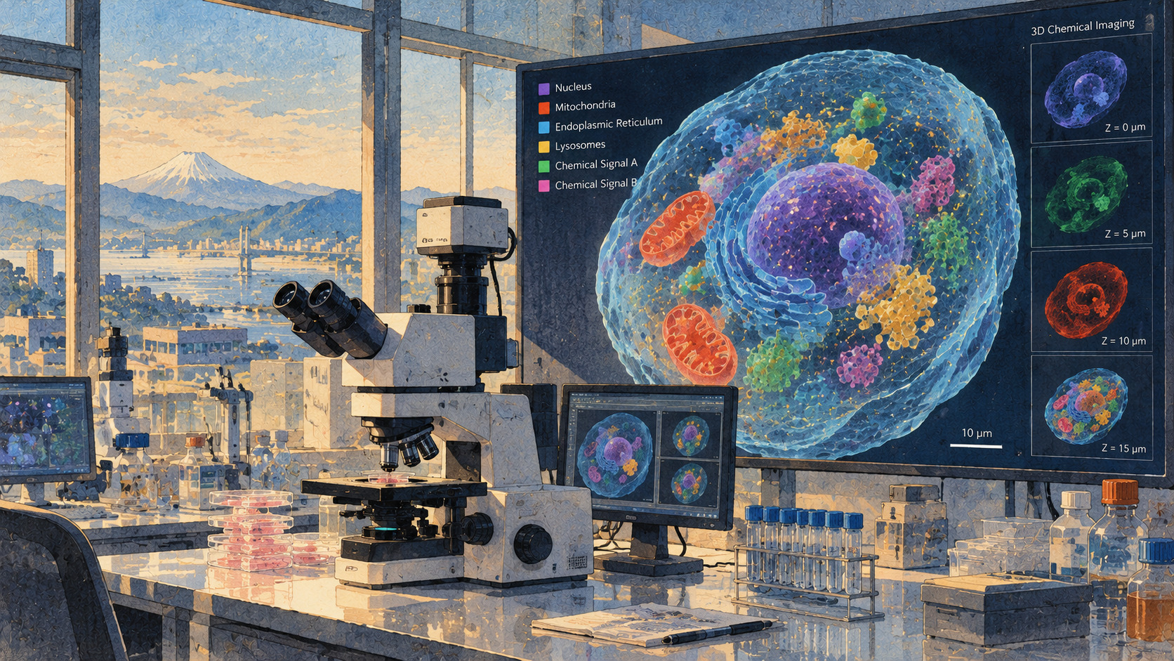
The breakthrough is not merely a faster picture. It is a moving, three-dimensional measurement with chemical contrast.
A living cell is not a still life
A cell’s interior is in constant motion. Tiny lipid-rich bodies travel through crowded spaces, chemical components gather and disperse, and movement along the depth of the cell can disappear when a microscope compresses everything into a flat image. A University of Tokyo team now reports a way to capture chemical information in complete three-dimensional frames fast enough to form a video—without first applying a dye. The result, published October 30 in Nature Communications, is a laboratory advance in how researchers can measure cellular activity rather than simply photograph it.
What changed: one complete volume every 52 milliseconds
The instrument produced 19.2 three-dimensional chemical images per second. Previous three-dimensional measurements with comparable mid-infrared photothermal tomography took less than 0.05 volumes per second, according to the researchers. That makes the new technique roughly 400 times faster within this particular class of methods. The figure should not be mistaken for a claim that all other microscopes are 400 times slower. One full volume now takes about 52 milliseconds, allowing motion that would have been averaged away in a slow scan to become a sequence of measurements.
Why the chemistry matters as much as the speed
Many powerful approaches to live-cell microscopy use fluorescent labels. Those labels can locate a selected protein or structure, but attaching them may require preparation and can sometimes perturb a biological system. Chemical imaging attempts something different: it draws contrast from the molecules already present. Mid-infrared absorption depends on molecular vibrations, so the wavelengths at which a sample responds provide information about its composition. This technique complements fluorescence methods rather than rendering them obsolete.
A two-color trick with light
Mid-infrared light excites molecular vibrations. Absorption creates a tiny photothermal change in a sample, altering its refractive index—the way it bends light. A visible-light probe detects that change. Comparing measurements with and without the infrared illumination isolates the chemically meaningful signal. Researchers then reconstruct the sample from these optical measurements; the result is not a conventional camera image of molecules, nor proof that every molecular species can be distinguished individually.
Eleven angles make the third dimension
The Tokyo system uses optical diffraction tomography. It gathers information under illumination from 11 different directions, then combines those views computationally into a volumetric image. A spatial light modulator rapidly changes the direction of the probe light, while improvements in signal quality avoid time-consuming averaging. In the paper’s terminology, the approach is video-rate mid-infrared photothermal optical diffraction tomography, or MIP-ODT. The engineering achievement lies in acquiring enough useful data for a volume without spending seconds on each reconstruction.
A test case: tracking the cell’s lipid droplets
To demonstrate a biological use, the group followed lipid droplets, structures that store lipids and participate in cellular metabolism. Lipid-associated optical signatures identified the droplets while the instrument tracked them in three dimensions. The observations permitted quantitative analysis of their trajectories, including behavior related to their locations inside the cell. A droplet moving deeper into a cell may appear nearly stationary in an ordinary two-dimensional view; volumetric tracking helps distinguish those paths.
A second mode adds a chemical spectrum
The team also demonstrated three-dimensional spectroscopic imaging at approximately 20 infrared wavenumbers per second. Wavenumber is a standard measure of position in an infrared spectrum, not a count of objects inside the cell. Sampling different parts of that spectrum supplies more chemical discrimination than imaging at one selected frequency. There are trade-offs among speed, spectrum and imaging conditions; neither demonstration means that a complete chemical inventory of every cell can already be recorded in real time.
A 350-year story of looking deeper
Optical microscopes opened microscopic biology to scientific examination in the seventeenth century. Later advances in staining and fluorescence let investigators pick out particular structures and molecules; confocal and modern computational methods added depth. Infrared spectroscopy developed along a separate path, revealing chemical bonds through their characteristic absorption. Connecting that kind of molecular signature with fast, three-dimensional live-cell observation has been a difficult intersection of optics, computation and biology.
The earlier Tokyo breakthroughs
The Ideguchi research group previously announced high-speed label-free mid-infrared filming in 2023 and a mid-infrared nanoscale-imaging achievement in 2024. These are related milestones rather than interchangeable specifications. High-speed two-dimensional imaging, very fine spatial resolution and fast full-volume chemical imaging each impose different optical and computational constraints. The 2026 paper is important because it removes a bottleneck in the third of those goals.
The Japanese researchers behind the method
The University of Tokyo names graduate students Masato Fukushima and Yusei Sugawara, Project Assistant Professor Keiichiro Toda and Project Researcher Shotaro Kawano in its Graduate School of Science, together with Professor Takuro Ideguchi in the Graduate School of Frontier Sciences and colleagues. Ideguchi also serves as a team director at the RIKEN Center for Advanced Photonics. The paper is titled “Video-rate volumetric chemical imaging via mid-infrared photothermal optical diffraction tomography.” Its DOI is 10.1038/s41467-026-78699-z.
Why researchers might want this—and what remains unproven
A promising longer-term question is how dynamic biomolecular condensates, involved in organizing proteins and nucleic acids, form and change inside cells. Some such processes are relevant to disease research. But the October study did not validate a clinical test, diagnose patients, show that a therapy works or establish long-term phototoxicity and cost for routine use. Further experiments will need to determine where the approach is robust and where fluorescence, Raman methods or other tools perform better.
From beautiful images to measurements
The most compelling consequence may be less spectacular than a colorful cellular movie: observations can become quantitative trajectories tied to chemical contrast. Researchers can ask how quickly a lipid droplet moves, where its course changes, and how its behavior varies inside the same cell. If the method proves transferable across materials and laboratories, it could give biologists an additional window onto life’s moving chemistry. For now, the result is a well-defined technical demonstration—and an invitation to test its reach.
Sources and verification
Research figures follow the university disclosures and journal record. Clinical applications remain prospective.