Night begins before sleep. It begins before the pineal gland releases its familiar darkness signal and before a conscious brain decides that the room has gone quiet. Deep in the hypothalamus, directly above the crossing of the optic nerves, a pair of nuclei smaller than a grain of rice changes its electrical language. Thousands of neurons that were comparatively talkative by day become quieter. The master clock does not switch off. It changes state.
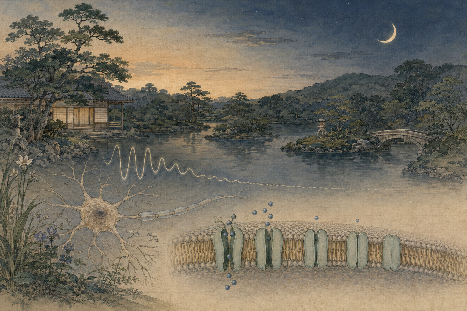
A Japanese-led team says it has identified one of the molecular gates that helps make that change. In neurons marked by the signaling peptide prokineticin 2, or Prok2, G-protein-gated inwardly rectifying potassium channels—GIRK channels—carry a persistent current at night. Potassium flows across the cell membrane, the inside of the neuron becomes more negative, and firing is suppressed. The cell acquires an electrical night.
The image is simple: open a gate, let positive charge leave, quiet the neuron. The science is not. The suprachiasmatic nucleus, or SCN, contains many neuronal types, each with its own peptides, receptors and timing. Multiple families of sodium, calcium, chloride and potassium channels shape their daily voltage rhythms. Glial cells also participate. Molecular clocks tick inside individual cells while the cells continually reset one another. To identify one channel in one named population and then connect it to an animal’s behavior required a ladder of evidence—from electrical recordings and single-cell gene tests to fluorescent anatomy, targeted genome editing, whole-network measurements and weeks of mouse movement.
What “night” means in this paper
The headline needs a boundary. The researchers did not discover the molecule that creates darkness, sleep, dreaming or melatonin. They identified a mechanism that contributes to the nighttime electrical state of a particular population of neurons in the mouse SCN. “Night” is shorthand for low excitability: a more negative resting membrane potential, less readiness to fire and reduced spontaneous activity during the appropriate circadian phase.
That distinction matters because sleep and circadian time are related but not identical. Sleep pressure builds with time awake; the circadian system places windows of greater wakefulness and sleep propensity across the day. A mouse is nocturnal, yet its SCN neurons—like those of many diurnal mammals—are generally more electrically active during the biological day and quieter during the biological night. Downstream circuits interpret the clock’s timing differently: the same daytime SCN signal can help suppress locomotion in a mouse while supporting daytime organization in a human.
The experiment also distinguishes night from dark. Researchers use “zeitgeber time” when animals are kept on a light–dark schedule and “circadian time” when external cues are removed. In constant darkness there is no lamp transition to define night, but the animal’s clock continues to generate a subjective day and subjective night. A mechanism that still changes behavior under constant darkness belongs to the endogenous clockwork, not merely to a reflex caused by a dark room.
The evidence, rung by rung
The project brought together Kanazawa University’s integrative-neurophysiology group with collaborators at Hokkaido University and the University of Tokyo. Its authors combined expertise in circadian circuits, cellular electrophysiology, channel anatomy and targeted genome manipulation—disciplines that have to converge when a claim travels from a microscopic current to whole-animal timekeeping.
The paper by Jaehun Jung, Takashi Maejima, Miwako Yamasaki, Yusuke Tsuno, Mohan Wang, Yuichi Hiraoka, Masahiko Watanabe and Michihiro Mieda began with electrophysiology. In brain slices from mice, the researchers recorded identified Prok2 neurons across daytime and nighttime phases. The cells were more excitable by day and less excitable at night. A basal current with GIRK-like pharmacology contributed to nocturnal hyperpolarization, and its maintenance depended on G-protein signaling.
Electrical behavior alone does not identify the physical channel. The team therefore used immunofluorescence to look for channel proteins and single-cell reverse-transcription PCR to read messenger RNA from individual neurons. GIRK1 and GIRK3 emerged as the predominant subunits in Prok2 cells. GIRK channels are tetramers—four protein subunits assembled around a potassium-selective pore—so the evidence points to a GIRK1/GIRK3-containing complex rather than a lone molecule acting in isolation.
The decisive test was causal. Using an in-vivo genome-editing strategy, the team expressed Cas9 and a guide RNA in Prok2-producing neurons to disrupt GIRK3 selectively. That choice avoided a whole-body knockout, which would alter GIRK signaling in many brain regions and make the behavioral result difficult to locate. In the edited Prok2 cells, normal nocturnal hyperpolarization was diminished. The day–night rhythm of excitability flattened because night had become too electrically day-like.
The change then appeared at larger scales. SCN network activity was altered. In a light–dark cycle, nocturnal mice normally begin moving soon after the lights go out; edited mice waited roughly three hours. In constant darkness, controls maintained a period close to 24 hours, while the edited animals’ activity rhythm ran at 24.5–25.1 hours. On a double-plotted actogram—the barcode-like chart chronobiologists use—their activity band drifted later day after day.
| Evidence | What was measured | What it supports |
|---|---|---|
| Patch-clamp recording | Membrane voltage, firing and basal current in identified Prok2 neurons | Prok2 neurons are more excitable by day; a GIRK-like, G-protein-dependent current helps hyperpolarize them at night. |
| Immunofluorescence | Channel-subunit proteins in tissue | GIRK1 and GIRK3 are present in the relevant cell population. |
| Single-cell RT-PCR | Subunit messenger RNA from individual Prok2 neurons | The same cells carry the molecular instructions for the predominant subunits. |
| Cell-specific editing | GIRK3 disruption in Prok2 neurons | GIRK3 is causally required for the normal nighttime electrical state. |
| Network recording | Coordinated activity across the SCN | A cell-level change propagates into altered clock-network dynamics. |
| Actograms | Movement in light–dark cycles and constant darkness | The channel mechanism affects activity onset and endogenous circadian period. |
A clock found by destroying it
The road to this ion channel began with experiments much larger and cruder than a single-cell edit. For centuries, biologists knew that living things anticipate the day. Plant leaves continued to open and close in darkness; animals kept daily schedules without clocks on the wall. In 1971, Ronald Konopka and Seymour Benzer gave the field a genetic handle when they isolated three fruit-fly mutants: one arrhythmic, one with a 19-hour period and one with a 28-hour period. All mapped to a single region of the X chromosome later known as the period gene.
One year later, two groups located a central mammalian timekeeper by removing it. Robert Moore and Victor Eichler reported that lesions in the suprachiasmatic region abolished the daily corticosterone rhythm in rats. Friedrich Stephan and Irving Zucker showed that bilateral SCN lesions permanently eliminated circadian drinking and locomotor rhythms. The paired nuclei sat directly above the optic chiasm—hence supra-chiasmatic—in exactly the kind of position that could receive environmental light information.
A lesion proved necessity but not that the SCN itself generated time. In 1990, Martin Ralph, Russell Foster, Fred Davis and Michael Menaker performed the experiment that made the pacemaker vivid. They destroyed the recipient hamster’s SCN, then transplanted a tiny SCN graft. Rhythmic behavior returned. More remarkably, the restored period followed the donor. Tissue from a mutant hamster with a short intrinsic day imposed its short day on the recipient. The graft carried time.
1971 — Konopka and Benzer link altered daily periods in fruit flies to mutations at one genetic locus.
1972 — Independent rat lesion studies identify the SCN as necessary for major circadian rhythms.
1990 — Transplanted hamster SCN tissue restores rhythmicity and imposes the donor’s circadian period.
2002 — Prokineticin 2 is identified as a rhythmic signaling output of the SCN.
2017 — The Nobel Prize recognizes the molecular feedback mechanism built from period and allied clock genes.
2026 — GIRK1/GIRK3 is tied to nocturnal hyperpolarization in Prok2 neurons and to behavioral timing.
Genes can count time, but neurons must announce it
The great molecular-clock discoveries explained how a cell can sustain a near-24-hour oscillation. In the canonical loop, the proteins CLOCK and BMAL1 activate genes including Period and Cryptochrome. PER and CRY proteins accumulate, enter the nucleus and inhibit the machinery that produced them. As they are modified and degraded, inhibition lifts and another cycle begins. Interlocking loops add stability, phase and temperature compensation.
Jeffrey Hall, Michael Rosbash and Michael Young won the 2017 Nobel Prize in Physiology or Medicine for establishing the logic of this feedback system in fruit flies. Mammalian homologues turned the principle into a clock distributed through the body. Liver, muscle, lung, skin and many other tissues contain cellular clocks. The SCN is “master” not because it owns every oscillation, but because its light-entrained network coordinates the rest.
A transcriptional loop, however, is invisible to the next neuron. The SCN must translate the chemical state of genes and proteins into output: electrical spikes, calcium signals, neurotransmitters, peptides and patterns of secretion. That translation is two-way. The molecular clock changes ion-channel expression and function; membrane voltage and calcium feed back into clock-gene transcription. Time is not passed along a one-direction assembly line. It circulates between nucleus, membrane and network.
This is why the new study matters. It occupies the conversion point between “what time the cell’s genes say it is” and “what electrical message the cell sends.” A clock that oscillates internally but cannot alter its firing is like a watch with moving gears and no hands.
The gate with a misleading name
GIRK stands for G-protein-gated inwardly rectifying potassium channel. Every word carries history. The pore is selective for potassium. It is controlled by G proteins that are released downstream of G-protein-coupled receptors, or GPCRs—the enormous receptor family through which neurons sense neurotransmitters and neuromodulators. And it “rectifies” because intracellular magnesium and polyamines block outward current more strongly at positive voltages, giving the channel a preference for inward current under certain experimental conditions.
The name can mislead when applied to a resting neuron. Potassium concentration is much higher inside a cell than outside it. Around the usual resting membrane potential, opening a GIRK channel can therefore allow net potassium efflux. Positive charge leaves; the voltage moves closer to the potassium equilibrium potential; the inside becomes more negative. The neuron is hyperpolarized and less likely to reach the threshold for an action potential. A channel classified as an inward rectifier can still produce the outward potassium current that quiets a cell.
GIRKs were established in the 1990s as direct effectors of inhibitory GPCR signaling. When a ligand activates an appropriate receptor, the heterotrimeric G protein separates functionally, and its beta-gamma subunits bind the channel together with the membrane lipid PIP2. The pore opens rapidly without the long relay of making a new protein. In the heart, GIRK1/GIRK4 channels help parasympathetic acetylcholine slow the pacemaker. In the brain, combinations involving GIRK1, GIRK2 and GIRK3 carry inhibitory signals from receptors for GABA, opioids, serotonin, adenosine and other transmitters.
The 2026 result adds a new context: a tonic—or sustained—G-protein-dependent GIRK current that appears specifically at night in Prok2 neurons. Yet the upstream switch remains unknown. The paper identifies the gate and establishes that G-protein signaling holds it open, but it does not identify which GPCR and which ligand deliver the nightly command. That missing receptor is the next mystery and potentially the more selective therapeutic handle.
- An unidentified nighttime signal activates, directly or indirectly, a GPCR pathway in a Prok2 neuron.
- G-protein beta-gamma signaling and PIP2 favor opening of GIRK1/GIRK3-containing channels.
- Potassium conductance pulls the membrane voltage toward a more negative level.
- The neuron becomes less likely to fire action potentials.
- Its changed activity alters communication within the SCN network and the phase and period of behavior.
Prok2: the clock’s broadcast signal
Why study Prok2 neurons? Because prokineticin 2 has long looked like one of the SCN’s ways of broadcasting time. In 2002, Michelle Cheng and colleagues reported that Prok2 messenger RNA oscillates strongly in the SCN, responds to light entrainment and acts as a clock-controlled output molecule. Delivering the peptide at night suppressed rat locomotor activity; mice deficient in Prok2 later showed weakened behavioral rhythms. Animals lacking its receptor, Prokr2, retained cellular timekeeping in the SCN but lost precision and coordination in activity and body-temperature rhythms.
Prok2 therefore sits at a useful junction. It is a secreted peptide, but “Prok2 neuron” also labels a defined cellular population whose electrical activity can be studied and manipulated. The cells do not merely release a clock product. Their voltage and calcium rhythms contribute to the network state from which timed output emerges. Kanazawa researchers created knock-in mice that allow Prok2 neurons to be genetically accessed and, in 2023, recorded their calcium rhythms in freely moving animals.
The peptide’s history also warns against a simple wiring diagram. Prok2 and its receptors occur in multiple brain and body systems. SCN Prok2 cells form diverse subpopulations and mingle with neurons identified by vasopressin, VIP and other peptides. A signaling molecule can couple cells inside the SCN, carry output beyond it, and influence light-dependent sleep–wake behavior. The clock is not a command neuron with one wire. It is a parliament whose members speak overlapping chemical dialects.
There was already more than one way to make electrical night
Calling GIRK “the night channel” would erase a substantial literature. SCN neurons change voltage through the balance of many conductances. A daytime sodium leak involving NALCN promotes depolarization. Voltage-gated sodium and calcium channels support firing. Calcium-activated BK potassium channels help reduce nighttime firing; mice lacking the BK-channel gene show elevated nocturnal activity and weakened rhythms. Two-pore-domain potassium channels provide background leak currents: TRESK and TASK-3 have each been linked to nighttime hyperpolarization and stable responses to light.
These findings are not contradictions. Earlier experiments often studied the SCN as a whole or used global knockouts. The new paper asks about a named subpopulation and a receptor-linked channel. Different neurons can use different combinations, and the same neuron can use more than one brake. One conductance may set resting voltage, another shape each spike, another respond to calcium, and GIRK connect an extracellular signal to sustained inhibition.
The “bicycle” model of SCN excitability captures the division of labor. A depolarizing wheel turns by day as sodium conductance rises. A hyperpolarizing wheel turns by night as potassium conductances become dominant. The gears overlap and compensate. Removing one component may flatten voltage rhythms without completely abolishing behavior because other cells and channels preserve a network-level oscillation. Conversely, a modest change in a strategically placed population can shift phase or period even when the molecular clock keeps ticking.
| Conductance | Established role in SCN timing | How GIRK differs |
|---|---|---|
| NALCN sodium leak | Supports daytime depolarization and repetitive firing. | GIRK supplies an opposing, hyperpolarizing potassium route at night. |
| BK potassium channel | Shapes action potentials and helps suppress nocturnal firing. | BK is calcium- and voltage-activated; GIRK is linked directly to GPCR/G-protein signals. |
| TRESK / TASK-3 | Background K+ currents help set nocturnal resting state and light responsiveness. | The 2026 study resolves GIRK function specifically in Prok2 neurons. |
| GIRK1 / GIRK3 | Night-specific tonic current in Prok2 neurons; affects network activity, phase and period. | The upstream nightly receptor and ligand remain unidentified. |
The three-hour delay and the longer day
The behavioral results describe two related but distinct properties. The three-hour delay after lights-out is a phase or activity-onset defect under an entrained light–dark cycle. The mouse still encounters a 24-hour environmental schedule, yet begins its active phase late. The 24.5–25.1-hour rhythm in constant darkness is a period defect: with no daily light reset, the internal cycle itself takes longer to repeat.
Those phenotypes give the paper relevance to human circadian disorders, but not equivalence. Delayed sleep–wake phase disorder makes it difficult for people to fall asleep and wake at socially desired times. Non–24-hour sleep–wake rhythm disorder causes sleep timing to drift around the clock when the endogenous period cannot remain aligned with the day. The edited mice show features conceptually reminiscent of delay and period lengthening. They do not model the full human diagnoses, and the study did not test sleep architecture, human GIRK variants or patients.
Even “activity onset” must not be casually translated as “bedtime.” Mice are nocturnal, so lights-off marks the expected start of running, exploration and feeding. Human sleep timing reflects the SCN, homeostatic sleep pressure, light exposure, behavior, social schedules and many downstream arousal and sleep circuits. A three-hour mouse locomotor delay is evidence that the clock network changed, not proof that GIRK3 deletion causes a three-hour sleep delay in people.
Why a direct GIRK drug would be difficult
Ion channels are attractive drug targets because small molecules can alter their function quickly. GIRK channels, however, are widely used. In the brain they participate in inhibition, reward, analgesia, mood, cognition and responses to opioids and alcohol. Related GIRK complexes regulate heart rate. A drug that opens or closes GIRKs everywhere could change much more than circadian phase.
Subunit selectivity helps but does not solve the problem. GIRK3, encoded in humans by KCNJ9, is not unique to the SCN. A compound would also need to reach the correct brain region at the correct time, influence the relevant GIRK1/GIRK3 complex and avoid altering other neuronal populations. Chronotherapy—timing a medicine to biological phase—adds another variable: the same intervention could advance a clock at one circadian time and delay or disrupt it at another.
The unknown upstream GPCR may offer a better path. If Prok2 neurons receive a distinctive nighttime signal through a receptor enriched in that population, modulating the receptor could be more selective than blocking the potassium pore throughout the body. But first researchers must identify the ligand, prove its rhythm, map the receptor’s distribution and show that manipulating it shifts behavior without unacceptable effects.
Existing human treatments provide a reality check. Carefully timed light can reset the clock through retinal input to the SCN. Melatonin or melatonin-receptor agonists can help align some disorders when used appropriately. These approaches act on known entrainment systems; their effect depends strongly on timing. A GIRK-based therapy would need to demonstrate an advantage in efficacy, precision or tolerability over those tools.
The experiments that should come next
The first task is to find the nightly hand on the gate. Researchers can screen GPCRs expressed in Prok2 neurons, block candidate transmitters, monitor G-protein activity across circadian time and ask whether a receptor’s deletion reproduces the GIRK3 phenotype. Because GIRK current was tonic, the signal may be continuously present at night, or the receptor–channel complex may itself change sensitivity with the clock.
The second task is to separate channel composition from cell identity. Does GIRK1 partner only with GIRK3 in these neurons, or do smaller GIRK2-containing populations matter? Is GIRK1 required to traffic or conduct? Rescue experiments—restoring GIRK3 selectively after deletion—would strengthen causality. Temporally controlled editing or acute pharmacology could distinguish an adult signaling role from developmental compensation.
The third task is to map the network consequence. Which SCN cells receive altered Prok2-neuron output? Does GIRK3 deletion change peptide release, calcium phase, synchrony or the spatial wave of SCN activity? Does the molecular PER2 rhythm lengthen in the same cells, or does electrical mis-timing reshape the network period without changing each intracellular oscillator? Multi-day imaging and cell-type-resolved recording can answer those questions.
Then comes translation. Human stem-cell-derived SCN-like neurons, postmortem expression maps and genetic association data can test whether a comparable GIRK1/GIRK3 mechanism exists in people. Naturally occurring KCNJ9 variants could be examined for chronotype or sleep-timing effects—but only with large cohorts and careful control for light, work schedules and ancestry. A channel conserved in humans is not automatically used by the human SCN in the same circadian way.
- Identify the upstream GPCR, ligand and reason activation is night-specific.
- Rescue GIRK3 selectively and reproduce the phenotype with independent editing strategies.
- Measure molecular-clock phase, peptide release and network synchrony alongside voltage.
- Test whether the mechanism is shared across nocturnal and diurnal mammals.
- Confirm GIRK1/GIRK3 function in human SCN-like cells and human tissue.
- Develop cell-, subunit- and time-selective modulation before considering clinical trials.
A discovery about silence
Neuroscience is naturally drawn to activation. A neuron flashes, a circuit lights up, an animal moves. Inhibition looks like absence. Yet biological timing depends as much on the organized production of quiet as on the production of spikes. Without a low electrical night, the contrast of the daily signal shrinks. The network has less room to announce morning.
The Kanazawa-led study gives that quiet a molecular door. GIRK1 and GIRK3 assemble in Prok2 neurons. A G-protein-dependent signal opens the pore at night. Potassium conductance lowers the voltage. Firing subsides. When GIRK3 is removed only from those cells, night becomes electrically incomplete; the network changes, the mouse starts late and its internal day lengthens.
That sequence is compelling because it crosses scales without pretending they are the same. A protein is not a behavior. A membrane voltage is not a diagnosis. A mouse actogram is not a human life. But a rigorous chain can connect them, one experiment at a time.
The larger history of circadian biology has moved through such translations. A fly mutation turned time into a gene. A brain lesion turned time into a place. A transplanted nucleus showed that tissue could carry a period. Molecular feedback loops explained how cells count hours. Calcium imaging and cell-specific genetics revealed a network of many clocks. The new work asks how one part of that network makes night legible in electricity.
Reporting notes and principal sources
This article is based on information available through August 8, 2026, 6:00 a.m. Japan Standard Time. The core experiments were performed in mice of both sexes. “Creates night” refers to nocturnal hyperpolarization and reduced excitability in Prok2-expressing SCN neurons; it does not mean that GIRK channels alone create darkness, melatonin secretion, sleep or subjective night. Comparisons with human delayed sleep–wake and non–24-hour disorders are mechanistic context, not diagnoses established by the study.
- Kanazawa University: August 7 research announcement, figures, genome-editing design and behavioral results
- The Journal of Neuroscience: Jung et al., “GIRK Channels Regulate Circadian Rhythms of Excitability in Prokineticin 2 Neurons…”
- DOI record: 10.1523/JNEUROSCI.0260-26.2026
- Nature / PubMed: 2002 identification of Prokineticin 2 as an SCN circadian output molecule
- PNAS: Prokr2 is essential for coordination of circadian behavior and physiology
- Scientific Reports: in-vivo calcium rhythms in SCN Prok2 neurons
- The EMBO Journal: single-cell SCN populations and the Prok2–Prokr2 signaling axis
- Brain Research / PubMed: Moore and Eichler’s 1972 SCN-lesion experiment
- PNAS: Stephan and Zucker’s 1972 SCN lesion study of drinking and locomotion
- Science: transplanted suprachiasmatic nucleus determines circadian period
- Caltech / PNAS: Konopka and Benzer’s 1971 clock mutants of Drosophila
- Nobel Prize: 2017 award for molecular mechanisms controlling circadian rhythms
- Journal of Physiological Sciences: review of ion channels controlling SCN excitability
- PLOS ONE: BK channels and spontaneous firing rhythmicity in the SCN
- Nature Communications: TRESK and nocturnal SCN dynamics
- Chronobiology International: TASK-3 channels, SCN electrical rhythms and light entrainment
- Nature Reviews Neuroscience: physiology and signaling of GIRK channels in the brain
- Trends in Pharmacological Sciences: GIRK channels as disease targets and the challenge of selective modulation
- U.S. National Heart, Lung, and Blood Institute: types of human circadian rhythm disorders
- U.S. National Heart, Lung, and Blood Institute: light, melatonin and treatment context