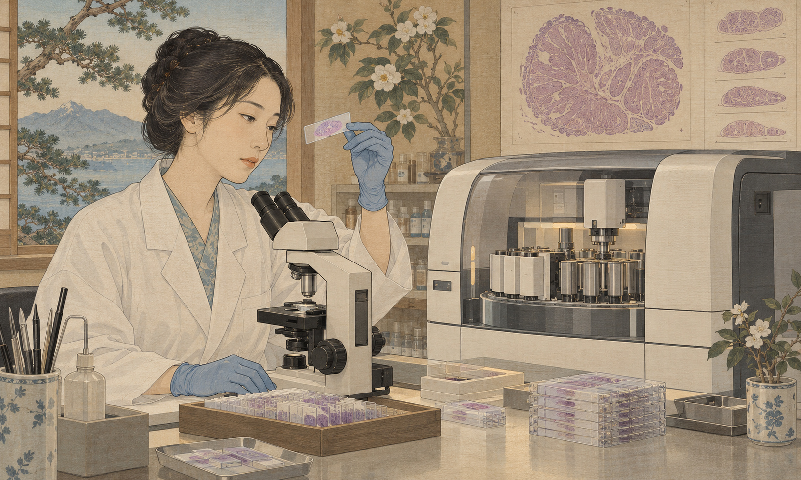
Before a pathologist can interpret a tissue sample, someone must make it possible to see. The work includes cutting an exceptionally thin section, spreading it out and transferring it onto glass. Path One, announced on September 8, addresses this physical preparation stage of pathology.
Akita University says the device grew from needs identified by its clinical staff. Its September 7 announcement identifies a collaboration involving Path Imaging, Institute of Science Tokyo, the Akita Industrial Technology Center and local financial institutions.[1][2]
The story is about making the material that diagnosis depends on. Understanding that role is essential: automation of tissue-section preparation is different from an automated diagnosis, and its significance is best judged in the laboratory workflow it is intended to support.
Why a thin section matters
The National Cancer Center Japan explains that pathological examination can establish whether a lesion is cancerous and inform treatment decisions. Tissue examination allows the pathologist to assess tissue architecture as well as the appearance of individual cells.[4][5]
In its general explanation, the manufacturer describes sections approximately 3–5 micrometers thick. Four micrometers equals 0.004 millimeters. This provides a sense of scale; it is not a published statement of the device’s full adjustable thickness range or its manufacturing tolerance.[2]
At that scale, cutting is only part of the task. The resulting section still has to be handled and placed on a slide. Looking at the blade alone would miss the sequence of transfers that turns a slice of material into something ready for subsequent preparation and examination.
Four connected operations
The Akita Industrial Technology Center describes four automated operations: sectioning, spreading or flattening the section, picking it up onto glass, and storage. The center says its contribution included analysis of complex water-flow control.[3]
| Stage | Purpose |
|---|---|
| Sectioning | Cut a thin section from a paraffin block. |
| Flattening | Spread the section after contraction during cutting. |
| Pickup | Transfer the section onto a glass slide. |
| Storage | Place it in storage for subsequent work. |
This outline follows the published descriptions.[2][3] It does not mean that one instrument performs everything from tissue collection and processing through staining, scanning and interpretation.
The water-flow work is a revealing detail. Connecting operations requires control over the material between them, as well as at the cutting point. That is an inference about the engineering challenge, not an independently observed account of the machine’s internal design.
A development history measured in years
The company’s account traces its involvement with the 北東北ナノ・メディカルクラスター研究会, a northern Tohoku nanomedical research group, to 2016. Requests to ease technicians’ specimen-preparation workload led to development beginning in December 2019. The September 2026 announcement therefore followed almost seven years of development.[2]
The university’s release records Path Imaging’s establishment in October 2014 and identifies its headquarters in Yokohama and an office in Kakunodate, Semboku, Akita. The clinical problem was rooted in Akita, while the collaboration extended beyond the prefecture.[1]
This history offers a useful view of how a clinical requirement becomes an engineering project. A hospital can identify the work it needs help with; a device developer must translate that requirement into repeatable operations. An industrial research center can contribute analysis that a clinical team would not ordinarily undertake itself.
The resulting machine is thus also a product of connections maintained over time. Its development illustrates the importance of having a place where medical staff can explain a practical difficulty to people capable of examining it as a design problem.
Digital pathology still begins with physical material
Path Imaging’s existing product information lists digital slide scanners including the Fino-SH/Fino-WH and UH-5S/UH-50L. Those instruments handle imaging; Path One addresses an earlier preparation stage. The company’s scanner specifications should not be mistaken for specifications of the sectioning device.[6]
The distinction is especially relevant when pathology is discussed in terms of digital images and artificial intelligence. An image originates in a physical specimen. Improving one part of that chain can be useful without making every other part automatic.
The industrial technology center expresses an expectation that more uniform specimens could support better performance in pathology AI.[3] That is a development rationale, rather than a reported comparison establishing improved diagnostic accuracy. The quality of a prepared section and the performance of a particular diagnostic system need to be assessed separately.
For readers, the appropriate questions are concrete: what aspect of specimen quality became more consistent, how was it measured, and did the change improve a downstream task? A general promise of better AI cannot substitute for those answers.
A longer concern with specimen quality
Standardizing the handling of pathological material predates this device. The Japanese Society of Pathology published guidance for handling pathological tissue specimens for genomic research in March 2016 and released a second edition in July 2025.[7]
On its separate guidance page for genomic clinical practice, the society places specimen quality assurance at the foundation of reliable cancer genomic medicine.[8] This is broader professional context, not an endorsement or evaluation of Path One.
That history helps explain why preparation technology remains consequential even as analytical methods become more sophisticated. A new instrument should be considered within the complete quality-management process: which task does it stabilize, and which tasks still depend on other equipment, procedures or professional judgment?
Automating one stage cannot retrospectively change how a specimen was collected or handled earlier. Nor does a uniform section alone establish that all subsequent tests will perform as intended. Defining the contribution narrowly makes it possible to evaluate it meaningfully.
What would demonstrate a practical benefit?
A laboratory assessing automation would need to look beyond the speed of a single successful operation. Preparation, loading, checking, repeat work and cleaning all belong in an assessment of workload. These are evaluation considerations, not measured results reported here for Path One.
Throughput and staff time are also different quantities. A machine may process material for a long period, while its value to technicians depends on how much direct attention it requires. If time is released, another question follows: what necessary work can staff do with it?
Quality deserves the same practical approach. Performance across the kinds of samples encountered in routine work matters more than the appearance of one successful demonstration. Staff also need a way to identify a problem and decide what happens next. An automated sequence can change the location of human judgment without eliminating the need for it.
These considerations are particularly important when discussing workforce benefits. The existence of a product does not establish a measured reduction in overtime or staffing pressure. Those outcomes emerge from how equipment is integrated into actual operations.
The patient’s perspective
For a patient waiting for a result, the relevant clock covers the entire examination. The National Cancer Center Japan explains that pathological results can take several days to several weeks.[4] A change to one preparation stage cannot be converted directly into a promised reduction in an individual patient’s waiting time.
Path One nevertheless brings attention to a part of medicine that is easy to overlook: skilled preparation before interpretation. Its announcement is a concrete result of an Akita clinical need being carried into product development.
The next useful account will be about everyday operation—usable specimen quality, the work required from technicians and the effect on the laboratory as a whole. Those measures will show how far the device answers the practical need that started its development.
Sources and references
- Akita University and partners: September 7, 2026 announcement
- Path Imaging: Path One release, September 8, 2026
- Akita Industrial Technology Center: development support, September 11, 2026
- National Cancer Center Japan: pathological examination
- National Cancer Center Japan: tissue examination
- Path Imaging: digital slide-scanner product information
- Japanese Society of Pathology: specimen-handling guidance publication history
- Japanese Society of Pathology: specimen quality in genomic medicine