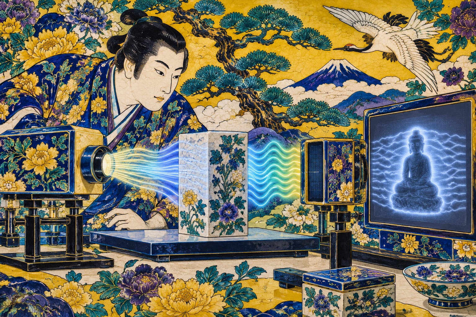
The breakthrough begins with an apparent contradiction. A shadow seems to be where information disappears. In the right optical system, however, its bright and dark edges can preserve information that a conventional intensity image cannot show: how far a wave has advanced or fallen behind as it passes through an object.
Shusaku Mikami of Chiba University’s Graduate School of Science and Engineering demonstrated the idea with an international team that included Professor Katsuhiko Miyamoto of Chiba, Assistant Professor Seigo Ohno of Tohoku University and Assistant Professor Adam Vallés of the Autonomous University of Barcelona. Their paper, “Terahertz phase-sensitive imaging via spiral-phase filtering and single-pixel detection,” appeared in Optics Express on September 2, 2026.
A Region Between Radio and Light
Terahertz radiation occupies a difficult and useful stretch of the electromagnetic spectrum between microwaves and infrared light. It can pass through many dry, nonmetallic materials that block visible light, including paper, fabrics, polymers and ceramics. Different molecules and solid-state structures can also respond characteristically at terahertz frequencies, making the band attractive for spectroscopy as well as imaging.
Those advantages come with boundaries. Metals strongly reflect terahertz waves, and water absorbs them. Spatial resolution is limited by wavelengths far longer than visible light unless specialized near-field methods are used. Terahertz radiation is non-ionizing, unlike X-rays, but that physical distinction is not by itself a complete safety or clinical validation for a particular instrument.
The useful measurement is not merely how much radiation survives a sample. Intensity records attenuation. Phase records the wave’s position in its oscillation cycle—effectively how its journey has been delayed. A sample that barely changes intensity can still alter phase because of its thickness or refractive index. Measuring both can therefore reveal boundaries or variations that amplitude alone leaves inconspicuous.
Phase has traditionally demanded more elaborate instrumentation. Terahertz time-domain spectroscopy measures the electric-field waveform as it changes in time. Interferometry compares a sample beam with a reference beam. Both approaches can be highly informative, yet synchronization, path stability, scanning and physical footprint can complicate an instrument. The new study asks whether phase can first be converted into a pattern that an intensity detector knows how to record.
How the Apparatus Makes Phase Cast a Shadow
The experiment starts with a highly monochromatic, frequency-tunable terahertz source. After the beam encounters the object, lenses guide it through a 4f system. In that classic optical arrangement, the central Fourier plane organizes the image by spatial frequency rather than by ordinary position. Placing a filter there changes how those spatial components recombine into an image.
The team uses a spiral phase filter at that Fourier plane. The filter twists the wavefront according to angle, producing what is often called an optical vortex. When the components are transformed back into an image, boundaries are emphasized. Under controlled conditions they look less like a uniform outline than like relief illuminated from one side: a bright edge paired with a dark one.
That directional “shadow effect” is the information channel. Its orientation and contrast depend on the phase entering the system. The researchers observed the bright-dark pattern reverse when the handedness of the vortex was reversed. They also changed the recovered phase distribution by rotating the spiral phase plate. Those deliberate reversals help separate a phase-dependent signal from an accidental blemish in the image.
| Component | Job | Why it matters |
|---|---|---|
| Tunable monochromatic source | Illuminates the sample at a selected THz frequency | Brings phase sensitivity to a continuous-wave-style measurement |
| 4f optical system | Exposes a Fourier plane for spatial filtering | Provides the stage for controlled wavefront processing |
| Spiral phase filter | Turns phase structure into directional edge contrast | Creates measurable shadow clues |
| Single-pixel detector | Records sequentially encoded total signals | Reconstructs an image without a detector array |
“Intensity Only” Needs a Careful Translation
The institutional announcements accurately describe a reconstruction from intensity images. That phrase can nevertheless invite the wrong mental picture. The method does not take an arbitrary photograph and discover phase afterward through software alone. Before detection, the optical system has deliberately converted phase structure into intensity structure. Computation then decodes those structured measurements.
Nor does one uncontrolled shadow uniquely reveal every possible phase object. The result depends on known filter settings, the measurement model and multiple phase-sensitive observations. The experimental achievement is the construction of an invertible trail: phase influences the spiral-filtered edge pattern in a controlled way, and that pattern supplies enough constraints to estimate the spatial phase distribution.
That distinction is more than semantics. It identifies where complexity has moved. Conventional interferometry encodes phase by comparing two optical paths. This design moves part of that work into Fourier-plane filtering and numerical inversion. Eliminating a reference arm could reduce one source of alignment sensitivity, but the alternative still needs stable characterization of its source, filters, modulation and reconstruction pipeline.
A Twenty-Year Trail From Microscopy to THz
The shadow principle did not begin in 2026. In 2005, Alexander Jesacher and colleagues at the Medical University of Innsbruck reported “Shadow Effects in Spiral Phase Contrast Microscopy” in Physical Review Letters. A spiral element in a microscope’s Fourier plane enhanced edges, while control of its central phase broke the symmetry and produced a rotatable, relief-like shadow. The paper demonstrated the effect on a low-contrast human cheek cell.
Spiral phase contrast subsequently developed as a way to emphasize edges and visualize phase objects. A separate line of work attacked the shortage of affordable, sensitive terahertz detector arrays. In 2008, researchers demonstrated a compressed-sensing terahertz camera built around a single detector pixel and a sequence of masks. Instead of collecting every image coordinate at once, the system inferred an image from known spatial codes.
By 2020, single-pixel detection and spiral phase contrast had been combined in visible-light experiments for edge detection of amplitude and phase objects. The Japanese-Spanish team has now carried the phase-sensitive shadow concept into terahertz imaging and, according to the universities’ announcement, used that sensitivity in the THz region for the first time to reconstruct spatial phase from intensity data.
The project also has a visible development path in Japan. Conference records from 2024 and 2025 list work by members of the same group on broadband terahertz spectroscopic single-pixel imaging and terahertz edge enhancement. The new paper is therefore better understood as the convergence of three mature ideas—Fourier optics, structured light and computational imaging—than as an isolated trick.
2005 · Spiral-phase microscopy produces controllable shadow effects.
2008 · Compressed sensing enables a single-pixel terahertz camera.
2020 · Single-pixel spiral phase contrast is demonstrated in visible light.
2024–2025 · The Chiba-led group reports THz single-pixel and edge-enhancement work.
2026 · Spatial THz phase is reconstructed from spiral-filtered intensity patterns.
Why a One-Pixel Camera Is Not a Paradox
A one-pixel camera does not ask one detector to know where every photon arrived. It asks a sequence of known questions. The illumination or transmitted field is modulated with one spatial pattern after another. The detector returns a single total for each pattern. Because the system knows every pattern it displayed, an algorithm can solve backward for the two-dimensional distribution most consistent with all of those totals.
This trade can be attractive in spectral regions where focal-plane arrays are expensive, insensitive, small or difficult to operate. A sensitive point detector may outperform each element of an available array. Computational imaging can also select efficient measurement bases and, when the object is sufficiently sparse or structured, use compressed sensing to reduce the number of observations.
But sequential acquisition is a real cost. If the object moves while many patterns are being measured, the reconstruction can blur or fail. Fewer measurements can improve speed while degrading fidelity. Resolution, signal-to-noise ratio and acquisition time compete with one another. Neither the university summaries nor the accessible publication record establishes a production-line frame rate, field performance or minimum detectable defect for the new configuration.
Where Better Phase Contrast Could Matter
Nondestructive inspection is the most immediate application class. A phase map can respond to small differences in thickness or refractive index across polymer components, composites, coatings, adhesive layers, paper products or packaged devices. If attenuation is nearly uniform, the delayed wave may still expose an internal boundary. In principle, that adds a second contrast mechanism to a conventional transmission image.
A tunable source also opens a path from single-frequency morphology toward spectroscopic imaging. Scanning frequency could test whether a feature has a material-specific response as well as a geometric one. Yet the 2026 paper is a method demonstration, not a certification study for a particular aircraft composite, semiconductor line or civil structure. It reports no universal defect-detection rate, false-alarm rate or factory inspection time.
Biological measurement requires even more discipline in the language. Water’s strong terahertz absorption limits penetration into hydrated tissue, especially at depth. The same sensitivity may create useful contrast near a surface or in thin, controlled samples. The researchers list biological measurement among future possibilities, but this experiment involved no patients and did not establish diagnostic accuracy, clinical utility or the safety profile of a medical device. A promising imaging mechanism is not a diagnosis.
The Tests That Must Come Next
- Speed: How many encoded measurements and how much time are required for one phase map?
- Accuracy: How closely does reconstruction match calibrated thickness and refractive index?
- Resolution: What smallest feature survives the wavelength, optics and sampling process?
- Robustness: Can the method tolerate vibration, humidity, rough surfaces, scattering and multiple layers?
- Tradeoffs: How quickly does fidelity decline as the number of patterns is reduced?
- Benchmarking: Does total cost and performance beat THz-TDS, interferometry and other computational methods?
Reflection geometry will matter as well. Transmission is convenient for a laboratory sample that can be placed between source and detector. A large structure or a human body usually demands access from one side. Reflections add surface roughness, multiple paths and phase wrapping to an already delicate inverse problem.
The team’s “first in the terahertz region” language should also be read at the claimed level: use of the phase-sensitive shadow effect created by spiral-phase filtering to recover THz phase. Japan.co.jp has not independently adjudicated every prior experiment worldwide, so the article attributes that priority to the research institutions rather than presenting it as a settled universal fact.
The listed authors are Shusaku Mikami, Yusuke Odagiri, Wataru Senzaki, Adam Vallés, Seigo Ohno and Katsuhiko Miyamoto. Chiba University says the work received support from four Japan Society for the Promotion of Science KAKENHI grants—JP23K23248, JP24KK0108, JP22K18979 and JP23K04567—and the Japan Science and Technology Agency FOREST program under JPMJFR2036, among other support.
Simplification by Moving the Work
Scientific instruments often become simpler not when complexity disappears, but when it is moved to a more manageable place. Digital photography shifted chemistry into electronics. Computational imaging shifts some lens and detector functions into known modulation and inference. Here, a task normally assigned to a reference beam is partly reassigned to a spiral phase element and reconstruction mathematics.
That may prove valuable. A system without an interferometer could be less vulnerable to some path-length and alignment problems. A single sensitive detector could avoid the cost of a large THz array. Frequency tuning could add material information. Whether those advantages survive outside a controlled apparatus will depend on calibration, acquisition speed, environmental stability and honest comparison with techniques that already measure the full terahertz field.
The enduring image from the research is therefore not one of effortless vision through matter. It is subtler: phase leaves a signature even after an intensity detector appears to erase it, provided the wavefront has first been asked the right optical question. The shadows around an edge are no longer a nuisance to suppress. Properly designed, they become evidence.
- Tohoku University, Sept. 4, 2026 — institutions, researchers, method, terminology and paper record.
- Chiba University CHIBADAI NEXT, Sept. 4, 2026 — research background, findings, researcher comment and funding.
- Mikami et al., Optics Express 34(18), 2026 — paper record and DOI 10.1364/OE.610381.
- Jesacher et al., Physical Review Letters, 2005 — the shadow effect in spiral phase contrast microscopy.
- Chan et al., Applied Physics Letters, 2008 — a compressed-sensing single-pixel THz camera.
- Liu et al., Optics Letters, 2020 — single-pixel spiral phase contrast.
- “Roadmap of Terahertz Imaging 2021” — historical and technical context for THz imaging, miniaturization and computational methods.
Editor’s note: This report uses material available through September 5, 2026. Applications described are technical prospects, not demonstrations of a commercial product, field inspection performance or clinical diagnosis. The priority claim is attributed to the research institutions.