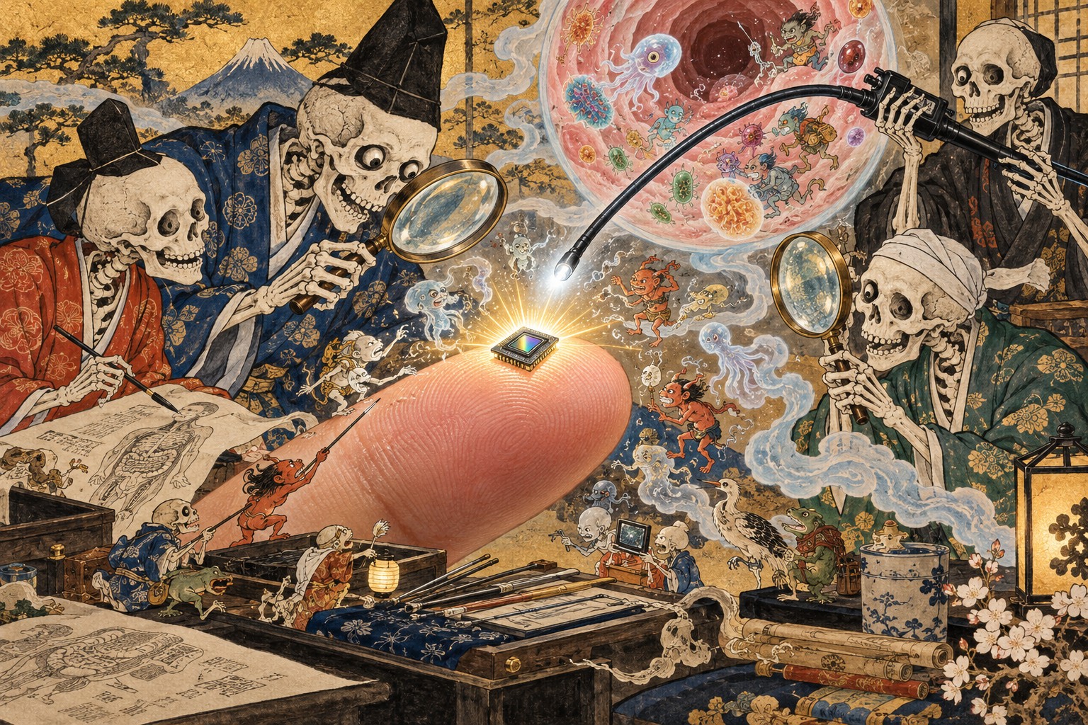
The breakthrough is a building block, not a proven medical breakthrough. What device makers can do with it is the next story.
A medical camera small enough to sit on a fingertip
The extraordinary fact about Sony’s latest image sensor is not simply that it measures one millimeter by one millimeter. It is that Sony says it has packed a digital camera sensor, its analog-to-digital conversion circuitry and the means to communicate over a long, extraordinarily thin cable into that footprint. In a conventional consumer camera, such details are hidden inside a spacious body. In an endoscope, every fraction of a millimeter at the tip can matter.
What Sony actually announced
On October 22, Sony Semiconductor Solutions Corporation said it would commercialize the IMX742, a back-illuminated CMOS image sensor intended for manufacturers of single-use medical endoscopes. Sample shipments are scheduled for November 2026. It is a component announcement, not the release of a finished bronchoscope, ureteroscope, diagnostic service or hospital-approved system. Sony claims an industry first for the combination of digital output and transmission through a five-meter cable among medical-endoscope CMOS sensors; that comparison is expressly based on its own research as of the announcement date.
The engineering inside a square millimeter
The IMX742 has 720 × 720 effective pixels, about 0.51 megapixels, on a 1.00 × 1.00 mm chip. Each pixel measures 1.00 micrometer across. Sony lists a back-illuminated structure, Bayer color filter, rolling-shutter and global-reset modes, and a six-pin wafer-level chip-scale package. The device supports 30 or 60 frames per second. These are consequential design choices, but not evidence that a finished medical camera will deliver a given clinical outcome: illumination, optics, image processing, packaging, power and the complete device all matter.
Why five meters can be as important as one millimeter
An endoscope has to travel from the distal camera tip through the insertion section and the operator’s handle to external equipment. A cable that carries delicate analog video can pick up interference along the route. Sony digitizes the image at the sensor and uses proprietary bidirectional communication to carry image and control information over the same cable, helping constrain the connection to six pins. Its five-meter figure comes from evaluation with AWG44 wiring in Sony’s test environment. It should not be read as a guarantee that every endoscope design, installation or electromagnetic environment will work at that distance.
What patients might gain — and what is not yet proven
Sony points to bronchoscopes and ureteroscopes as potential applications. A thinner imaging assembly may help designers navigate narrow spaces or accommodate other endoscope components; cleaner video and 60-fps output may aid real-time viewing. But a sensor by itself cannot prove a reduction in pain, complications, missed lesions or procedure time. Those questions need complete-system validation and, where applicable, clinical evaluation. Nor should “half a megapixel” be mistaken for a ready-made claim of diagnostic superiority.
The infection-control reason disposable endoscopes exist
Traditional reusable endoscopes are cleaned and disinfected or sterilized between patients in a process called reprocessing. The FDA has documented persistent difficulties with particularly complex duodenoscopes and has supported designs that facilitate or eliminate difficult reprocessing steps. Its findings do not establish that all reusable scopes are unsafe: proper reprocessing remains essential and reusable systems continue to have a major clinical role. Single-use devices trade some reprocessing burdens for new cost, supply-chain and waste challenges.
Japan’s long history of looking inside the body
Japan’s contemporary endoscopy industry rests on an unusually direct connection between physicians and optical engineers. In 1949, a University of Tokyo physician urged Olympus to develop a practical way to photograph the stomach. Olympus completed a prototype gastrocamera in 1950 and released the GT-I in 1952. The challenge then was fitting optics, a light source and film into an instrument that could enter the human body. Later fiber-optic systems and electronic video imaging transformed that achievement. Sony’s tiny digital semiconductor addresses a modern incarnation of the same problem: how to put useful vision into very little space.
Why the market opportunity is not just the sale of a chip
There is an industrial logic to Sony’s move. Camera sensors once associated with consumer photography now have applications in factory inspection, vehicles and medical equipment. For endoscope makers, the value of a reliable tiny component may lie in the system design it enables: distal-tip dimensions, cable routing, image electronics and potentially manufacturing scale. The announcement provides no IMX742 selling price, procurement volume, customer roster or revenue forecast. It therefore cannot substantiate a claim that Sony has already captured a particular share of the disposable endoscope market.
A difficult trade-off: infection prevention versus medical waste
Single-use equipment can simplify certain infection-prevention workflows, but disposable cameras, polymers, packaging and electronics create material and disposal burdens. The net environmental result depends on device construction, manufacturing, transport, reprocessing alternatives and local waste handling. Without a device-specific lifecycle analysis, it would be irresponsible to proclaim either single-use or reusable endoscopes universally greener. The industry will need to account for performance, safety, economics and waste together.
The crucial regulatory line
Sony explicitly cautions that the IMX742 is not itself a medical device and has not been evaluated or approved for medical applications. It is supplied for incorporation into equipment such as medical devices. That means claims about regulatory clearance, hospital availability or clinical performance must refer to particular finished products, not to this chip announcement. The planned November date is for samples, not clinical deployment.
What to watch after the announcement
The next meaningful milestones will be sample delivery, integration by endoscope manufacturers, announced finished systems and substantiated comparisons under realistic imaging conditions. It will also matter whether the advertised digital link works across varied hardware configurations; whether engineers can maintain adequate illumination and thermal performance at the tip; and how hospitals assess total ownership and disposal costs. Those are questions, not announced outcomes.
The larger meaning of a one-millimeter square
For decades, Japanese medical imaging engineers have tried to push clear vision deeper into the body while making instruments less intrusive. The IMX742 represents another technical step in that pursuit. A commercially useful digital image path within a square millimeter is striking. The responsible conclusion is narrower, and more interesting, than a promise of medical revolution: one of the world’s leading image-sensor makers has given endoscope designers a new building block. What they construct with it remains to be demonstrated.
Sources and verification
Based on Sony primary documentation, FDA records, Olympus archives and specialist interviews published by third parties. No original clinical testing or interviews were conducted.
- Sony Semiconductor Solutions, Japanese primary announcement (October 22, 2026)
- Sony Semiconductor Solutions, English release
- Sony: IMX742 product specifications
- EE Times Japan: engineer interview, October 22, 2026
- EE Times Japan: long-cable design and engineering context
- Olympus: birth of the gastrocamera
- Olympus: 1946–1959 company chronology
- US FDA: infections associated with reprocessed duodenoscopes
- US FDA: reprocessing of reusable medical devices