- When killing a cancer cell is only half the battle
- A newly published study, not a new approved treatment
- Why cancer-cell death can be immunogenic—or quiet
- The genetic material inside the cell’s power stations
- KRAS mutations offered an unexpected clue
- A drug triggers the alarm—and the cleanup crew
- Cytochrome c changes the fate of a dying cell
- The mouse result: two complete regressions among twelve animals
- A story that runs through Kyoto’s Nobel-winning immunology
- What a mitochondrial-DNA test would still have to prove
- The research question that has changed
When killing a cancer cell is only half the battle
Cancer drugs can destroy tumor cells and still leave a larger question unanswered: what happens after those cells die? A treatment may shrink a tumor without teaching the immune system to recognize the cancer that remains. A different kind of cellular death can leave a signal that recruits immune defenses. Understanding why these outcomes diverge is increasingly important as oncologists try to combine molecularly targeted drugs with immunotherapy.
An international team anchored at the National Cancer Center Japan, Kyoto University and Nagoya University has identified a consequential factor in that choice. Their peer-reviewed study finds that the abundance of mitochondrial DNA—and its balance with another mitochondrial molecule, cytochrome c—helps determine whether drug-induced cancer-cell death provokes an antitumor immune response.
A newly published study, not a new approved treatment
The paper, “Mitochondrial DNA Abundance Is a Determinant of Immunogenic Cell Death in Cancer,” appeared in Immunity on October 30, 2026 in the United States, October 31 in Japan. The participating institutions announced the results on November 2. Its senior corresponding investigator is Hiroyoshi Nishikawa, a professor associated with Kyoto University and the National Cancer Center Japan; Kosuke Tanaka, a specially appointed associate professor at Kyoto University’s Hakubi Center, is a co-first and corresponding author.
The study draws on cancer-cell datasets, mechanistic laboratory experiments and preclinical animal models. It does not report results from a human trial of the proposed three-drug combination. That distinction matters in a field where promising experimental biology can be mistaken for imminent medical treatment.
Why cancer-cell death can be immunogenic—or quiet
Ordinary apoptosis is a tightly managed form of cell death that can allow the body to clear damaged cells without a major inflammatory response. Immunogenic cell death is different: the dying cell releases or exposes signals that help stimulate immune recognition. The terms are not a ranking of good and bad death in every setting; apoptosis is necessary to normal physiology.
For a patient with cancer, however, the distinction may determine whether treatment does more than eliminate the cells it directly reaches. Dendritic cells must encounter and present tumor antigens, and killer CD8-positive T cells must then mount a meaningful response. Targeted drugs and checkpoint inhibitors can serve complementary functions, but the biological basis for successful combinations has remained incomplete.
The genetic material inside the cell’s power stations
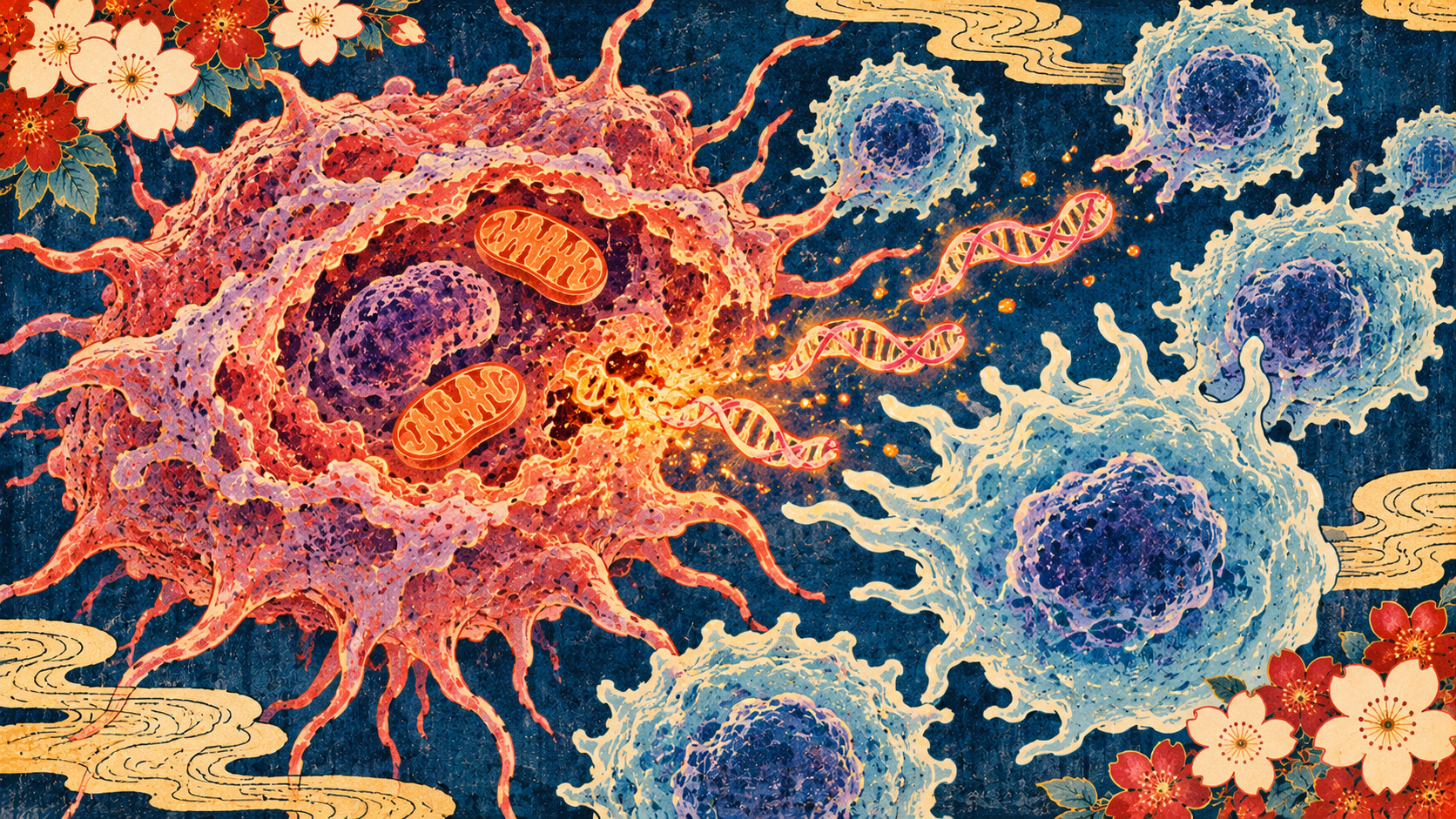
Mitochondria are often introduced as the cell’s energy-producing compartments. Less widely appreciated is that they retain their own DNA, separate from the genome housed in the nucleus. DNA is useful when contained within the right structure. When mitochondrial DNA spills into the surrounding cytoplasm, the cell can interpret its unexpected presence as danger.
One detection mechanism is the cGAS–STING pathway, which can activate type I interferon signaling and innate immune responses. The researchers explored whether the amount of mitochondrial DNA available to escape a damaged organelle was a critical constraint on this alarm system. The answer was not simply “more DNA means more immunity”: the pathway must function, and the context of cell death matters.
KRAS mutations offered an unexpected clue
When the researchers compared public cancer-cell-line datasets, they found that mitochondrial DNA content correlated particularly strongly with KRAS mutation status, rather than merely with the organ where a cancer arose. Elevated levels appeared across KRAS-mutant cell models associated with cancers including lung, colorectal, pancreatic and biliary tumors. The team linked the enrichment to oxidative stress and increased mitochondrial DNA replication.
KRAS is a major driver of cell-growth signaling and a longstanding target of cancer research. The study suggests that molecular features within a tumor—not only conventional labels such as “lung cancer” or “colon cancer”—may help researchers anticipate immune responses to therapy. It does not establish KRAS status alone as a validated clinical test for selecting a new treatment.
A drug triggers the alarm—and the cleanup crew
To probe the mechanism, researchers used drugs that inhibit MEK, part of a growth-signaling pathway downstream from KRAS. Blocking this pathway helped prompt mitochondrial DNA to move into the cytoplasm. Yet the same intervention also activated mitophagy, the cell’s machinery for removing damaged mitochondria. The cleanup process reduced the amount of DNA available to set off an immune alarm.
In laboratory experiments, suppressing mitophagy kept mitochondrial DNA release going and strengthened cGAS–STING and type I interferon signaling. The institutional release describes use of chloroquine as an experimental inhibitor of that recycling process. Cells with little mitochondrial DNA, or without functional STING, did not show the same response. This is an experimental finding, not advice to take chloroquine or to modify prescribed cancer therapy.
Cytochrome c changes the fate of a dying cell
The study’s second insight concerns cytochrome c, a mitochondrial protein whose release can activate the machinery of apoptosis. In cells with abundant mitochondrial DNA, targeted treatment plus mitophagy inhibition could support immune-stimulating death. But in some EGFR-mutant lung cancer cells with high cytochrome c, conventional apoptosis predominated. Manipulating cytochrome c levels altered that balance under the tested conditions.
More aggressive treatment was not automatically more immunogenic: under sufficiently strong stimulation, apoptosis could dominate and immune signaling weaken. This adds a counterintuitive dimension to combination therapy design. Maximizing immediate cellular destruction might not maximize the immune response that could help control surviving disease.
The mouse result: two complete regressions among twelve animals
In a KRAS-mutant colorectal cancer mouse model, adding an anti-PD-1 checkpoint antibody to a MEK inhibitor and a mitophagy inhibitor improved antitumor activity. The National Cancer Center Japan reports that tumors disappeared in two of twelve mice in the relevant experiment, and that dendritic cells and CD8-positive T cells contributed to the response. The effect was absent in low-mitochondrial-DNA tumors under the investigated conditions.
That two-of-twelve figure must never be described as a response rate in people. Mouse tumors and human cancers differ; the efficacy, dosing, safety and interactions of this approach in patients have not been demonstrated by the study. Even a striking preclinical result is a starting point for translation, not a treatment protocol.
A story that runs through Kyoto’s Nobel-winning immunology
The work enters a field shaped by discoveries made in Japan and abroad. In 1992, Tasuku Honjo and colleagues identified PD-1, later shown to act as a brake on immune T cells. James P. Allison’s studies of CTLA-4 revealed another inhibitory checkpoint. Their work helped establish the therapeutic logic of releasing those brakes, and the two scientists shared the 2018 Nobel Prize in Physiology or Medicine.
Checkpoint blockade has transformed care for some cancers, but many patients do not benefit or eventually develop resistance. Removing a brake cannot by itself guarantee that immune cells see or recognize a tumor. The new mitochondrial findings address a different part of the chain: what signals a treated tumor provides to help recruit immunity in the first place.
What a mitochondrial-DNA test would still have to prove
One potential application is to use mitochondrial DNA abundance as a biomarker for choosing future combinations. That possibility remains unvalidated clinically. Investigators would need a reproducible assay, thresholds that work across laboratories and tumors, and trials showing the marker predicts benefit beyond existing clinical information. Tumor heterogeneity, changes under treatment and immune-cell composition would all complicate interpretation.
Equally important are safety and comparative effectiveness. Drug combinations may add toxicity. Chloroquine’s role in an experimental model does not establish it as an appropriate oncology treatment. A meaningful clinical advance would require evidence that patients live longer, feel better or otherwise obtain measurable benefit—without unacceptable harms.
The research question that has changed
The central insight is about the quality of tumor-cell death. Scientists traditionally count how many malignant cells a drug kills. This study argues for measuring what those cells release, which molecular signals survive the cell’s own recycling mechanisms, and how the immune system receives them.
Its value today is a sharper biological explanation and a plausible set of experiments for the future, not a promise of a cure. The distance from mitochondrial DNA in a laboratory dish to an effective combination regimen in a clinic remains substantial. But the study makes one thing clearer: a cancer cell’s final messages may matter as much as the fact that it dies.
Sources and reporting method
Source-based reporting from November 2 institutional statements, the journal record and abstract, and historical documentation. Detailed experimental data are attributed to the institutions; the full journal article may require subscription access.
- Kyoto University — Research announcement, Nov. 2, 2026
- National Cancer Center Japan — Joint research announcement and methods
- Immunity — Original peer-reviewed paper (DOI)
- PubMed — Abstract and bibliographic record
- Nobel Prize — 2018 Physiology or Medicine background
- US National Cancer Institute — History of checkpoint treatment