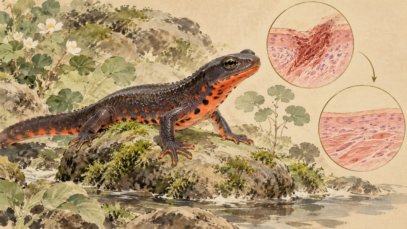
The breakthrough question is not just how to avoid a scar, but how tissue can resolve fibrosis already formed.
The scar that did not stay
A scar is often the price of survival. When a deep wound closes, the body lays down collagen-rich tissue that protects the damaged area, yet may leave stiffness, contracture or visible change. In a small amphibian native to Japan, researchers observed something that challenges the permanence of that bargain: scar-like fibrosis developed after skin transplantation, then diminished.
The study was published in Inflammation and Regeneration on August 1, 2026 and announced by Mie University and the University of Tsukuba on August 28. Science Japan returned attention to it on October 19. It was not a paper first published in October. The surprising finding is not merely that newts heal well. It is that their tissues appeared able to dismantle a fibrotic response after it had already formed.
A tiny transplant with a visible color difference
The animal was the Japanese fire-bellied newt, Cynops pyrrhogaster, whose dark back and red abdominal skin give scientists a convenient natural contrast. Under anesthesia, the team took a roughly 3-by-3-millimeter square of belly skin and sutured it into a similarly sized wound on the back. They compared grafts moved within the same animal with grafts from another animal of the same species.
This distinction matters. A graft from another individual exposes the recipient to foreign tissue and should provoke immune recognition. In humans, an allogeneic skin graft cannot normally provide a permanent substitute for a patient’s own skin because rejection becomes a problem. The researchers used the transplantation challenge to ask whether a highly regenerative animal avoids fibrosis altogether or manages to overcome it once triggered.
First the graft thickened. Then the fibrosis receded.
The foreign newt grafts did not visibly fall away. Instead, beneath the grafts, collagen-rich tissue and inflammatory cells accumulated. Histology identified fibrosis after approximately two weeks, maximum thickening around four weeks, and substantial regression by eight weeks. The external redness of the transplanted belly skin also faded as the area took on a more back-like appearance.
Crucially, this is not evidence that donor skin simply vanished. The study detected structural elements of transplanted tissue after prolonged observation, including at 60 weeks. It describes persistent integration alongside marked tissue remodeling. Nor was every aspect of inflammation eliminated: the animals developed a fibrotic reaction, but it did not remain at its peak.
An immune cell at the center of the puzzle
Immune cells are sometimes portrayed as agents of damage, but healing also depends on clearing debris, coordinating signals and resolving inflammation. The researchers tracked Iba1-positive cells, interpreted as macrophage-lineage cells. These cells accumulated in the graft during the pronounced fibrotic phase at four weeks, then became scarce when fibrosis had largely regressed by eight weeks.
The pattern was suggestive but correlation alone would not show that the cells mattered. So the scientists treated some animals with clodronate-loaded liposomes before transplantation, transiently depleting macrophage-like populations. The foreign grafts still formed fibrosis. What changed was the speed of its removal: thickened tissue persisted longer, including at the eight-week check.
What the intervention proves—and what it does not
The depletion experiment strengthens the case that early macrophage-lineage responses help fibrosis resolve. It does not establish a complete cellular pathway, identify a drug target for people, or prove that one precisely defined macrophage subtype independently carries out the cleanup. Clodronate liposomes are a research intervention affecting susceptible immune cells, not a demonstrated scar-reversing therapy.
The authors themselves caution that amphibian immune-cell classifications remain incomplete. Genetic lineage definitions are not as developed as those available for some mammals. They also could not determine how much of the outcome reflects the newt’s regenerative capacity and how much reflects its unusual response to transplanted tissue. The degree of donor–recipient immune mismatch was not fully characterized.
A lineage of research, from lost limbs to quiet immune work
People have long marveled at salamanders for regrowing limbs and other complex tissues. Modern regenerative biology has broadened the question: not only how new cells form, but what the injured tissue environment allows them to do. In 2013, James Godwin and colleagues reported that macrophages were necessary for successful axolotl limb regeneration; depleting them led to persistent fibrosis and disrupted regrowth.
Later work studied macrophages during regeneration of the newt lens and other structures. The 2026 Japanese skin-graft experiment extends that conversation in an important direction. Instead of simply asking whether a regenerative animal avoids scarring, it creates an immune challenge that induces fibrotic tissue and follows what happens as that tissue disappears.
Why plastic surgeons would care
Surgeons treating serious burns or traumatic skin loss have immediate priorities: keep patients safe, prevent infection and close the wound. A patient’s own skin is a cornerstone of reconstruction, but donor and recipient sites can still scar. A scar is not just an aesthetic issue. Excess collagen and remodeling can limit movement, cause discomfort and require further treatment.
Learning how a living organism resolves collagen-rich tissue could eventually inform that work. Yet a newt is not a small human. Skin structure, temperature, immune recognition and the duration of tissue remodeling differ. The research did not test a treatment in people, and it provides no evidence that human surgical scars—or fibrotic diseases of the lungs or liver—can be reversed by the same intervention.
Japan’s collaboration between the clinic and the field
The first author, Kento Hosomi, works in plastic and reconstructive surgery at Mie University. Coauthors include Chikafumi Chiba of the University of Tsukuba, an institution associated with regenerative-biology research. The project puts a clinician’s persistent question—why severe wounds leave lasting marks—beside the experimental tools of a biologist studying animals that regenerate.
Mie University’s department described this as its second newt-related research paper. Its public announcement and the peer-reviewed article, rather than private conversations with the researchers, provide the evidence for this report. The work illustrates how the most consequential medical questions sometimes begin by looking outside familiar mammalian model systems.
The significance of two, four and eight weeks
A useful shorthand for the study is two weeks to visible fibrosis, four weeks to its maximum, and eight weeks to its regression. Those numbers refer to the observed newt-graft model—not a prediction for the speed of human wound healing, and not a universal calendar for every injury in this species. By later observation, donor-derived skin structures remained detectable despite surface remodeling.
Some histological comparisons involved small numbers of animals, including figures identifying n=3 at four weeks and n=4 at eight weeks. The researchers report statistical tests and controlled interventions, but replication across larger cohorts, additional immune-cell markers and distinct injury conditions will be needed before the mechanism can be described as settled.
What comes next
Future experiments could identify which macrophage-lineage populations are active during regression, what signals they release, and whether enzymes that remodel the collagen matrix are involved. Another key question is whether immune tolerance to the graft, regenerative biology, or their interaction permits fibrosis to resolve. Eventually researchers might test whether related mechanisms can be detected in mammalian tissues.
Those are research directions, not announced clinical trials. The wonder of the Japanese fire-bellied newt is that it overturns an easy assumption: the body’s fibrotic response need not always be the final state. Understanding how this animal makes it temporary is a more durable story than claiming science has already found a cure for scars.
Primary research and documentation
The research was published August 1 and announced by the universities August 28; October 19 coverage was not a new experiment or publication. No human treatment has been established.
- 三重大学・研究成果発表(2026年8月28日)
- 筑波大学 TSUKUBA JOURNAL(2026年8月28日)
- Hosomi et al., Inflammation and Regeneration 46, 31 (2026), open-access full text
- Original peer-reviewed article DOI
- 三重大学医学部附属病院・形成外科、論文紹介
- Science Japan, October 19, 2026
- Godwin et al., Macrophages are required for adult salamander limb regeneration, PNAS (2013)