- The pressure we cannot put a gauge on
- A pressure sensor that is a tiny floating capsule
- Why the stiffness of a membrane changes the measurement
- What a 0.1–50-megapascal result actually establishes
- Reading a faint signal against the tissue's own glow
- Two older scientific ideas become a new platform
- From deep-sea life to questions about human disease
- The essential distinction: an optical experiment is not clinical imaging
- What the next experiments must show
The pressure we cannot put a gauge on
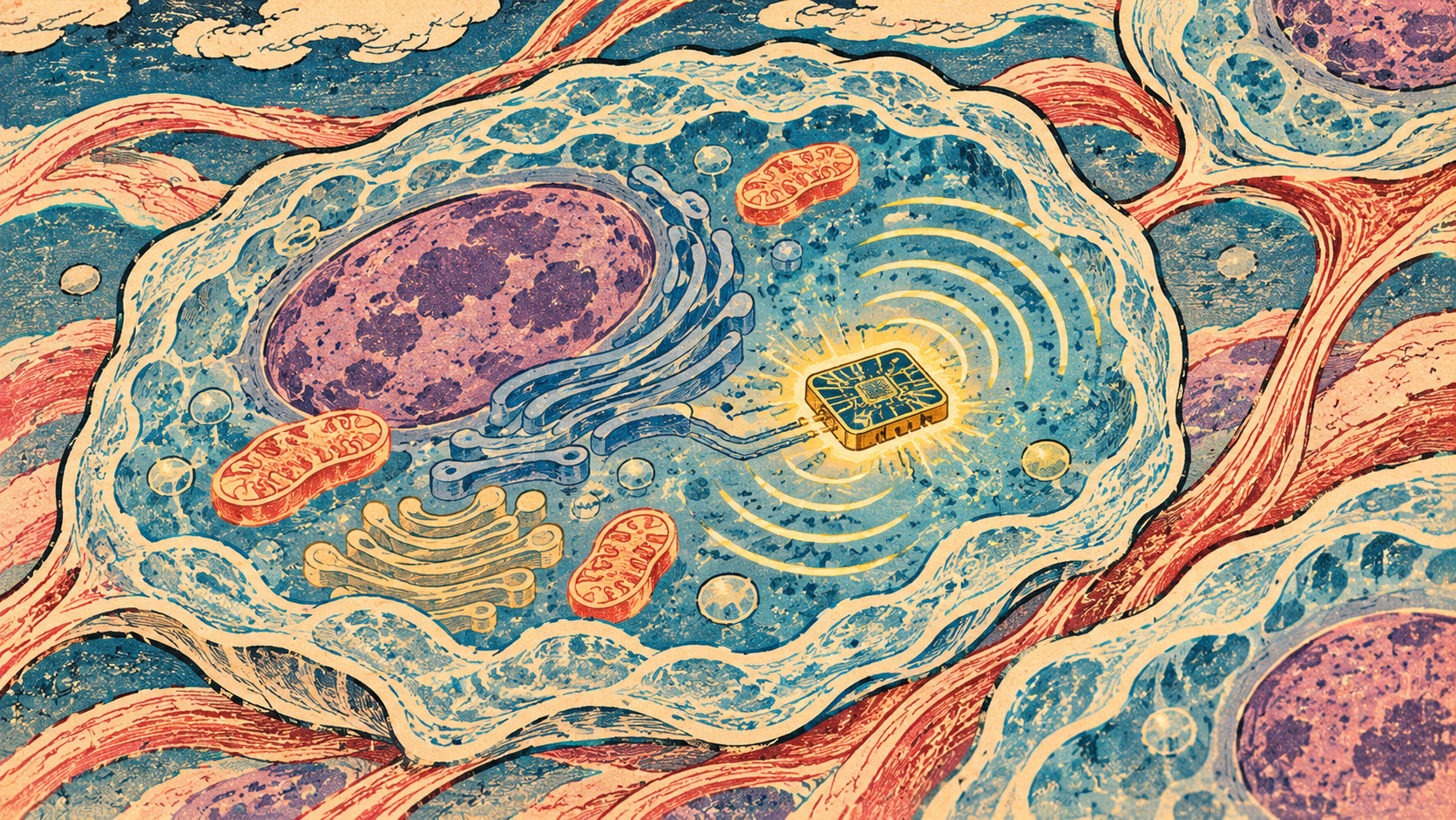
Inside living tissue, chemistry and mechanics are inseparable. Fluid surrounds cells, cartilage bears loads, and the heart moves against changing forces. Yet a pressure gauge that works in a pipe or laboratory vessel is a poor instrument for an aqueous space only fractions of a micrometer wide. Its presence may be larger than the thing scientists want to study.
A Japanese collaboration has approached the challenge with a material that reports pressure using light. Researchers at Institute of Science Tokyo, Tokyo University of Agriculture and Technology, and Kyushu University developed Pyr-PICsome, a polymer nanovesicle whose fluorescent response can be engineered by changing the stiffness of its membrane.
The underlying paper was published online August 15, 2026, and Japanese university announcements followed October 5. An English department feature appeared November 5, and Science Japan reported the research on November 20. The latest coverage is not a new November 20 experiment. The science is promising precisely because it addresses a hard measurement problem—but what has been demonstrated is narrower than the prospect of reading pressure deep inside living human tissue.
A pressure sensor that is a tiny floating capsule
The building block is a PICsome, short for polyion complex vesicle. It assembles in water from polymers carrying opposite electrical charges. The team held its diameter near 100 nanometers while altering chemical links in the membrane to make that membrane softer or stiffer.
Attached to the membrane is pyrene, a fluorescent molecule with a useful trick. When pyrene molecules remain apart, they emit mainly at a wavelength near 380 nanometers; when brought close enough together, they can form an excited-state pair, or excimer, with a different emission near 500 nanometers. Pressure changes the geometry of the membrane and thus the balance of those signals.
The ratio of the two light outputs provides a potential readout of hydrostatic pressure. Pyrene itself is not a new invention, and its poor water solubility has long complicated measurements in biological fluids. Binding it within a tunable, water-compatible capsule is the materials-engineering step that makes the method interesting. This is not a miniature chip implanted in a cell, and the capsule does not make a digital pressure reading by itself.
Why the stiffness of a membrane changes the measurement
The researchers tuned a membrane stiffness parameter from 7.3 to 39 piconewtons per nanometer without substantially changing capsule diameter. Those units describe a force-response measurement of the vesicle; they should not be confused uncritically with bulk Young's modulus of a conventional engineering material.
Under pressure, softer membranes let adjacent pyrene molecules approach each other more readily. The investigators report about fourteen times greater pressure sensitivity for the softest formulation than for the stiffest in the relevant fluorescence-ratio response. Stiffer variants, however, displayed useful changes in fluorescence lifetime.
The distinction gives the platform more than one possible measurement mode. One configuration favors how bright one signal becomes relative to another; another is suited to measuring how long the fluorescence persists. The word 'programmable' here means that chemists choose material properties during fabrication, not that software is steering an electronic sensor.
What a 0.1–50-megapascal result actually establishes
The reported hydrostatic pressure range is 0.1 to 50 megapascals. One megapascal equals a million pascals; 0.1 MPa is roughly atmospheric pressure, while 50 MPa is on the order of five hundred atmospheres. It is an appropriate scale for questions involving substantial water pressure, including marine environments.
It does not follow that the probe already resolves every biologically relevant pressure change. Pressures of interest in different tissues and diseases can be far smaller, and a large operating range is not equivalent to sensitivity at the low-pressure end. For a clinical application, the limits of detection, precision, temperature dependence, calibration and the practical location of the sensor would all matter.
A reader should also distinguish pressure from mechanical stress and strain. Hydrostatic pressure acts in all directions through a fluid; the force borne by a contracting muscle or the shear experienced by a moving cell is not necessarily captured by the same number. The best use cases will depend on the physical quantity researchers truly need to measure.
Reading a faint signal against the tissue's own glow
Biological samples are difficult optical environments. Proteins and other molecules may fluoresce under illumination, producing background light that obscures a small probe's signal. Measuring only light intensity also makes it hard to distinguish changes in probe concentration from actual changes in pressure.
The team reports excimer fluorescence lasting roughly 55 to 75 nanoseconds, compared with under five nanoseconds for much ordinary biological autofluorescence cited in the institutional release. A time-gated detector can ignore much of the early background and read the later emission; such behavior is attractive for fluorescence-lifetime imaging microscopy, or FLIM.
The scientists also tested the capsules in saline seawater and protein-rich culture media, reporting stability without aggregation or degradation under those evaluated conditions. That is a useful materials test, not a human biocompatibility trial or proof that the particles can be targeted, recovered and safely used in a patient for long periods.
Two older scientific ideas become a new platform
The story draws on two established research traditions. Chemists have used fluorescence changes to study molecular proximity and surroundings for decades. Pyrene excimer formation is a familiar example: the way its emission changes can indicate how molecules are arranged within a material.
Polyion complex vesicles come from a separate line of polymer and nanomedicine research: oppositely charged chains self-assemble into a membrane-enclosed structure in water. Such vesicles have been explored for biocompatible materials and delivery applications. Combining these strands allows membrane mechanics, rather than only dye chemistry, to shape the optical signal.
There is an institutional story too. Institute of Science Tokyo was established in October 2024 through the merger of Tokyo Institute of Technology and Tokyo Medical and Dental University. The current cross-university project includes Hayato Mizuno and Yasutaka Anraku at Science Tokyo, Yuki Akagi at Tokyo University of Agriculture and Technology, and Gaku Fukuhara at Kyushu University. The paper appears in ACS Applied Nano Materials as 'A Programmable Nanovesicle Platform for Megapascal Pressure Sensing.'
From deep-sea life to questions about human disease
The deep ocean offers one of the clearest scientific motivations. Microorganisms survive under enormous hydrostatic pressure, and researchers still have questions about how their membranes and molecular systems sense and adapt to those conditions. A water-soluble optical probe could help design experiments that were previously difficult to perform.
The biomedical possibilities are different and more demanding. Pressure dynamics are implicated in tissue swelling, joint and cartilage function, and aspects of cardiovascular and eye physiology. The university release highlights the possibility of future pressure imaging relevant to edema, glaucoma and musculoskeletal disease. It does not report that Pyr-PICsome has diagnosed glaucoma, replaced tonometry or mapped pressure in a patient.
The proposed next step is to add ligands on the capsule surface so that it can recognize selected cells or tissues. Even then, scientists would need evidence about delivery, local accumulation, optical access, clearance and whether the measurements reflect pressure rather than another changing property of the microenvironment. A sensor capable of functioning in a laboratory fluid does not automatically become a medical device.
The essential distinction: an optical experiment is not clinical imaging
It is tempting to describe a 100-nanometer capsule as a device that already maps pressure inside living tissue in real time. That would go beyond the clearest demonstrations in the published materials. The researchers evaluated pressure-dependent optical responses of engineered nanovesicles under controlled conditions and assessed their behavior in demanding aqueous media.
Using the method inside an organism requires several additional achievements: putting the probe in the desired place, bringing excitation light in and emitted light out, calibrating under biological conditions and distinguishing pressure from effects of temperature, acidity or probe concentration. The spatial resolution of a microscope is not automatically equal to the particle diameter.
University materials describe tissue-targeted and intracellular pressure sensing as development goals. The result therefore belongs in the category of enabling sensor technology, not established noninvasive clinical imaging. That qualification does not diminish the new design principle; it defines what future experiments must prove.
What the next experiments must show
The immediate contribution is the ability to design an optical pressure response through membrane stiffness. Instead of merely shrinking a conventional gauge, the researchers made a soft material itself the transducer. That opens a modular route toward different readout modes and perhaps eventually sensors responsive to several physical and chemical conditions.
The next evidence will need to come from calibrated tests in realistic tissues and cells, reproducibility across preparations, stability over time and comparison with independent measurement approaches. For a deep-ocean experiment, pressure, temperature and salinity must be controlled; for clinical translation, biocompatibility and the interpretation of results will be central.
A useful scientific instrument does not need an immediate medical application to matter. The value of Pyr-PICsome is that an invisible physical force can, under validated conditions, become a measurable change in light. Whether it will illuminate the pressure landscape of living tissue remains an important, unanswered question.
Sources, method and limitations
This independent explanation draws on the original ACS paper, October 5 Japanese university announcements, November 5 technical commentary and November 20 Science Japan coverage. We did not interview the researchers, replicate calibration tests or assess in-vivo or clinical performance. No validated clinical diagnosis by this probe has been established from the reviewed materials.
- 東京科学大学:2026年10月5日、日本語による公式研究発表 / Science Tokyo Japanese research release
- 九州大学:2026年10月5日、共同研究成果 / Kyushu University Japanese release
- 東京科学大学材料系:2026年11月5日、英語版解説 / Science Tokyo technical explainer
- Science Japan:2026年11月20日記事 / Nov. 20 coverage
- ACS Applied Nano Materials:A Programmable Nanovesicle Platform for Megapascal Pressure Sensing
- 東京科学大学材料系:研究成果の日本語解説 / Departmental Japanese explainer