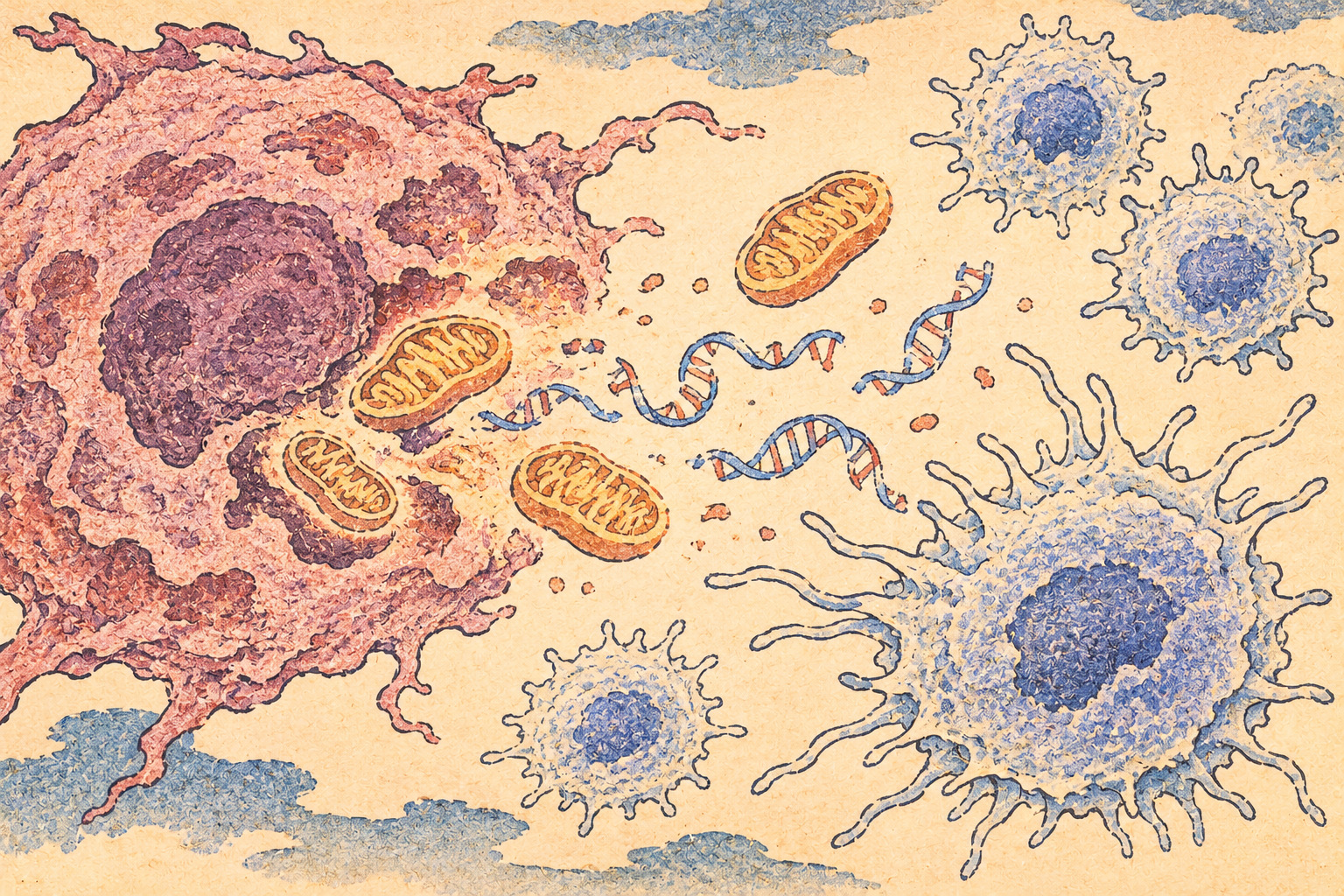
The moment after a cancer cell dies
For decades, success in cancer drug development has often been illustrated by a shrinking tumor. But the fate of a dying tumor cell is not the end of the story. Its molecular contents may be cleared away with little inflammatory response—or alert the immune system, potentially helping immune cells recognize other cancer cells that remain. The distinction could matter when targeted therapies and immunotherapies are used together.
Researchers from Japan's National Cancer Center, Kyoto University and Nagoya University have reported a mechanism that helps explain those different outcomes. In laboratory models, the balance between mitochondrial DNA and the protein cytochrome c helped determine whether targeted drug-induced cell death generated immune signaling. The findings appeared in Immunity on October 30, 2026, and were announced publicly in Japan on November 2.
What was discovered—and what was not
The new research is chiefly a mechanistic investigation involving cancer cell lines and mouse tumor models. It does not report a clinical trial showing that an experimental three-drug combination benefits patients. In one mouse experiment involving KRAS-mutant colorectal tumors, investigators observed stronger tumor suppression after combining a MEK inhibitor, a mitophagy inhibitor and an anti-PD-1 antibody; two of 12 animals had no detectable tumors at the reported endpoint.
That result is worth investigating, but it is not a two-in-twelve human cure rate. Animal models represent selected biological circumstances, and studies of small groups of mice do not establish human safety, effective dosing or clinical survival benefit. The journal article and Japanese research announcement should be read as a possible path toward future drug-combination research, not as instructions for medical treatment.
Mitochondria have a second identity
Mitochondria are usually introduced as the cell's energy-producing structures. They also carry their own DNA, separate from the much larger genome housed in the nucleus. In a healthy cell, the location of that DNA is tightly controlled. When mitochondrial DNA appears in the cytoplasm, it can be interpreted as a sign that something has gone wrong.
A cellular sensing route involving cGAS and STING can detect out-of-place DNA and initiate signaling associated with type I interferons. Such signals can help shape immune responses. The Japanese team found that releasing mitochondrial DNA, rather than simply possessing a large quantity of it, was important. Cells lacking STING did not show the studied response, underscoring the importance of intact sensing machinery.
Why KRAS drew the scientists’ attention
The investigators examined public cancer-cell-line datasets and found that mitochondrial DNA abundance correlated strongly with KRAS mutation status, rather than neatly tracking the tissue from which a cancer arose. The pattern appeared in cell lines representing cancers of the lung, colon, pancreas and biliary system. The team linked mutant KRAS signaling with increased reactive oxygen species and enhanced mitochondrial DNA replication.
KRAS is a familiar oncogene because mutations can keep growth-related signaling switched on. But the study's finding does not mean that every KRAS-mutated human tumor contains enough mitochondrial DNA to respond to the proposed treatment. Cell lines are valuable experimental models, not a substitute for the molecular diversity of cancers inside patients.
A cleanup process that suppresses the alarm
Cells regularly remove damaged mitochondria through a selective recycling process called mitophagy. In the researchers' models, MEK inhibition helped mitochondrial DNA enter the cytoplasm, but also promoted mitophagy. That cleanup reduced the amount of DNA available to activate immune signaling—a paradox in which a protective housekeeping process may blunt a therapeutic immune alarm.
The researchers used chloroquine for its reported ability to inhibit mitophagy in their experimental setting. By slowing mitochondrial removal, they increased the persistence of released mitochondrial DNA and the associated type I interferon response. Chloroquine has effects beyond mitophagy, however, and this experiment is not evidence that people with cancer should take it or add it to an existing regimen.
The quieter death pathway: cytochrome c
Cytochrome c is central to mitochondrial biology and can also help activate apoptosis, an organized form of cell death. The team observed that certain lung-cancer models with abundant mitochondrial DNA—associated with KRAS mutation or ALK and ROS1 fusions—could develop immune-associated signaling after combined treatment. In some EGFR-mutant lung-cancer cells with relatively high cytochrome c, apoptosis predominated.
Changing cytochrome c levels altered that balance in the laboratory. Stronger drug stimulation could also tip the system toward apoptosis with weaker immune signaling. These findings argue against a simplistic claim that killing more cells always generates more immunity. They do not imply that apoptosis is universally immunologically silent; context, cell type and surrounding immune conditions matter.
A history of taking the brakes off immunity
Modern cancer immunotherapy builds on many decades of fundamental science. Investigators learned that tumors can exploit molecular checkpoints that restrain T cells. James Allison's work on CTLA-4 and Tasuku Honjo's discovery of PD-1 helped create an approach that blocks these inhibitory signals. They shared the 2018 Nobel Prize in Physiology or Medicine, and checkpoint inhibitors have since changed treatment for multiple cancers.
But removing the brakes on an immune cell is not the same thing as providing a clear signal that a tumor should be attacked. Many patients do not respond to checkpoint therapy, and responses depend on an intricate interaction among tumor genetics, immune cells and tissues. The Japanese study addresses an earlier link in that chain: how therapy-induced tumor death might create the signals that make an immune response more likely.
The three-drug mouse experiment
One of the headline results came from KRAS-mutant colorectal tumors grown in mice. The combination of a MEK inhibitor, a mitophagy inhibitor and an antibody against PD-1 suppressed growth. Researchers reported no detectable tumors in two of 12 treated mice. They also implicated dendritic cells, which present information about threats to the immune system, and CD8-positive T cells, which can kill targeted cells.
Equally telling was the negative result: tumors with low mitochondrial DNA did not show the same enhanced response. That contrast supports the study's mechanism while highlighting limits to generalization. The research release does not establish a universal treatment response rate, a commercially available drug combination or a timeline for a human trial.
Can mitochondrial DNA become a biomarker?
The most practical long-term question is whether measuring mitochondrial DNA abundance—possibly alongside cytochrome c and DNA-sensing competence—could help predict which tumors might benefit from particular combinations. A useful biomarker would have to be reliable across patient biopsies, disease stages, drug treatments and laboratory methods. It would also have to add information beyond the biomarkers clinicians already use.
Those hurdles are not minor. Cancer tissue includes immune cells, blood vessels and multiple tumor subpopulations, none of which is recreated perfectly by a culture dish. A correlation seen in cells does not itself provide a clinical diagnostic threshold. No validated patient-selection test emerges from this announcement, and the combination's safety and effectiveness in humans remain open questions.
A research direction, not a prescription
Japan's Agency for Medical Research and Development supported aspects of the project, including a program directed at future treatments for KRAS-mutant cancers. Its international relevance lies in a different way of framing therapy: rather than counting dead tumor cells alone, researchers may need to understand what those cells signal to the surrounding immune system.
The unresolved task is formidable: repeat the mechanism independently, establish how broadly it operates in human tumors, test interactions and toxicity, and determine whether any improved immune response translates into longer lives or better quality of life. For now the scientific contribution is precise but preliminary. A tiny genome inside mitochondria has offered a clue about why cancer cells can die in very different immunological ways.
Sources and reporting method
Desk research based on the November 2 joint Japanese research announcement, the published paper details and authoritative historical accounts from the US National Cancer Institute and Nobel Prize. No original interviews, independent experiments or clinical verification were conducted.
- National Cancer Center Japan, Kyoto University and Nagoya University, joint release, November 2, 2026
- Tanaka et al., Mitochondrial DNA Abundance Is a Determinant of Immunogenic Cell Death in Cancer, Immunity (October 30, 2026)
- US National Cancer Institute, history and clinical development of immune checkpoint inhibitors (July 2026)
- Nobel Prize official summary, 2018 Physiology or Medicine Prize to Honjo and Allison
- National Cancer Center research institute, 2026 announcement index