Take one grain of salt from the kitchen and place it on black paper. To the unaided eye it is a white point. Under modest magnification and light raking across its side, planes and angles emerge; the crystal throws light back. Sugar has a different outline. Dissolve either in water, let the drop dry and the original grain does not simply return. A new congregation of crystals grows at the receding edge. In minutes, the subject has changed from “a white speck” into a record of matter finding order.
The Microscopic World Observation Photo and Video Contest, launched July 23 by Tokyo-based KaraSeed, asks people across Japan to collect transformations like this from home, school, park and workplace. Entries close August 31. Participants observe familiar materials—leaves, water, sand, fabric, food, insects or hair—through a microscope and submit a photograph or video with a comment explaining what they noticed, found interesting, felt or wondered. No specialist knowledge is required.
Entrants do not need the organizer’s microHunter product. Smartphone, desktop and mobile microscopes of any brand or type are eligible. Two winners may each choose one microHunter kit from the official store. According to the launch announcement, selection will consider not only beauty but the observer’s point of view and the realization or discovery produced by looking. That leaves open the possibility that the best question, rather than the loudest color, will prevail.
A Sales Promotion—and a Doorway to Observation
The commercial setting should not be blurred. KaraSeed, established in Fuchu, Tokyo, in 2020, describes its business as product sales, manufacturing and importing and serves as microHunter’s authorized Japanese distributor. The lens family was developed by Shanghai-based QingYing E&T and optical researcher Lee Cui with microscope enthusiasts. TIME named QingYing’s related iMicro C one of its Best Inventions of 2022. KaraSeed’s own product material says the broader series had supplied about 100,000 units in more than 70 countries by May 2025.
This is therefore neither a public science prize nor a society-run, peer-reviewed award. It is a campaign operated by a company that sells the prize. Its decision not to require its own hardware nevertheless matters. An aging school microscope, a rival mobile lens and a conventional desktop instrument enter on equal stated terms. By beginning with “look” rather than “buy,” the promotion becomes an open observation project rather than a closed product ecosystem.
The July 23 launch release does not identify named judges, divisions, a results date or a projected number of entries. Applicants should read the official rules before submitting, particularly provisions on copyright and promotional use, privacy, permission for minors and image processing. If the project continues, publishing entry totals, reasons for selection, judging expertise and the educational uses of submitted work would make it more transparent and more valuable.
Robert Hooke Publishes a “New Visible World”
Compound microscopes appeared in Europe around 1600, although the identity of a single inventor remains disputed. The decisive step was not merely making an instrument. It was transferring what appeared in that instrument into a form another person could examine and trust. In 1665, with the support of the Royal Society in London, Robert Hooke published Micrographia.
Fleas, the point of a needle, plants and a thin slice of cork expanded across large pages in precise engravings. The Royal Society describes it as the first fully illustrated book devoted to microscopy. In its preface Hooke announced a “new visible world.” He compared the small compartments in cork to monastic rooms—cells—and the word eventually became the name of biology’s basic structural unit.
Those famous plates were not automatic camera records. Hooke combined fragments seen at different focal depths through trained drawing and engraving. From its beginning, a microscope image was a collaboration among optics, specimen, observer and recording technique. A smartphone image is no different. What appears on its screen is not nature untouched, but one optical system’s representation of nature.
The Cloth Merchant Who Saw “Little Animals”
After reading Hooke, Antonie van Leeuwenhoek of Delft pursued a different design. A cloth merchant accustomed to magnifiers used to inspect thread, he made extraordinarily small single lenses rather than compound microscopes. One lens sat between brass plates; a specimen was mounted on a pin and brought into focus by screws. The device was uncomfortable, but it could produce a relatively clean image.
The Science Museum Group records that he made more than 500 lenses, with some instruments magnifying about 200 times. From 1673 he sent observations to the Royal Society, describing blood cells, protozoa, sperm and living bacteria as minute “animalcules.” A merchant outside a university opened microbiology through glass, patience, writing and reproducible illustration. That history resonates with a 2026 contest that does not make credentials an entry requirement.
Magnification Is Not Resolution
Making an image larger is not the same as separating finer detail. Magnification describes enlargement. Resolution describes whether two closely spaced points can be distinguished as two. Enlarging a blurred photograph until it fills a display does not reveal a new hair or cell wall. A smartphone’s digital zoom cannot create detail absent from the optical image.
In 1873, German physicist Ernst Abbe expressed the resolution of a light microscope in terms of the wavelength of illumination and the light-gathering ability of the lens. Roughly 0.2 micrometers became the familiar practical boundary for conventional visible-light microscopy. A stable stage, correct focus, suitable illumination and a well-prepared specimen may produce more information than a product’s headline maximum magnification.
Electron microscopy used much shorter wavelengths to go beyond the optical wall. Ernst Ruska built what the Nobel committee describes as the first modern electron microscope in 1933; a commercial mass-produced instrument followed in 1939. Later, fluorescence and confocal methods selected particular structures inside cells. The 2014 Nobel Prize in Chemistry recognized super-resolved fluorescence techniques that worked around Abbe’s limit. A household lens and nanoscopy have profoundly different capabilities, but both begin by designing the optical method around the thing one wants to know.
Japan’s Century: From JOICO to Image Analysis
Nippon Kogaku K.K., now Nikon, was established in 1917 to build optical instruments domestically. In 1921 it invited German engineers, including Heinrich Acht, to transfer advanced design knowledge. Japanese technicians ground, assembled and adjusted miniature lenses by hand, and in 1925 the company released the JOICO microscope. The name came from “Japan Optical Industry Company.” Its maximum magnification of 765 times was advanced for its period, although international conditions prevented mass production.
In 1976, Nikon’s CF—Chromatic Aberration Free—system changed the design logic by correcting color aberration independently in objective and eyepiece lenses. In 2026, the Japanese Society of Microscopy recognized both JOICO and CF as “Microscope Heritage.” The path from hand-polished glass to live-cell observation, drug discovery, pathology, in vitro fertilization and AI-assisted image analysis crosses a century of Japanese precision industry and medicine.
By coincidence, on July 25—the date of this edition—the Tokyo Photographic Art Museum opens Nikon’s exhibition The Microscopic World: Illuminating the Future of Life, marking the company’s 101st year in microscopy. Through August 23 it presents the historic JOICO, documents, Nikon Small World and NIKON JOICO AWARD images, and applications across science, medicine and industry. Admission is free on opening day, Nikon’s foundation anniversary.
Fifty Years of the Micrograph as an Artwork
Nikon Small World began in 1975 to recognize people working in photography through the light microscope. In 2011 it added Small World in Motion for movies and time-lapse work. An independent expert panel judges originality, informational content, technical proficiency and visual impact. A researcher’s cell, an enthusiast’s insect and a polarized crystal can meet on the same plane as scientific information and visual form.
Microscopic photography contains a productive tension. Beauty draws an audience closer, yet the processing used to create beauty can push evidence further away. Fluorescence and electron micrographs may not possess “natural” colors visible to a human eye. Pseudocolor is a legitimate translation that distinguishes structure or molecule, but it can mislead when presented as though it were ordinary vision.
Nature Portfolio’s image-integrity standards say images should be minimally processed, that adjustments should generally apply to the entire image and that pseudocoloring and nonlinear changes should be disclosed. The U.S. Office of Research Integrity treats a scientific digital image as numerical data and advises preserving the original file. A summer contest is not a journal manuscript. Even so, noting “false color added,” “focus stack combined” or “cropped” gives a beautiful image more credibility.
The Smartphone Turns an Eyepiece into a Shared Screen
A traditional microscope asked one observer to look with one eye, then draw the view or carefully attach a camera. The smartphone combines sensor, display, recorder, editor and network. Align a small accessory lens over its camera and a family or classroom can watch the same moving organism, pause it, compare frames and share the result. That social change can matter more than raw magnification.
Cheap microscopy is not the achievement of one company. In 2014, a Stanford team published the Foldscope, an origami-inspired instrument made from paper and a small spherical lens. The research demonstrated rugged, low-cost brightfield, darkfield and fluorescence designs intended for education and resource-limited field use. In Scotland, the EnLightenment research initiative delivered more than 500 low-cost smartphone microscopes to over 100 secondary schools, reaching thousands of students aged 12 to 14 and demonstrating the platform’s educational and public-engagement potential.
Portability carries limitations. Automatic camera switching, autofocus, vibration, shallow depth of field, simple illumination, a narrow field and image compression vary by phone and affect the result. At higher magnification, a tiny movement throws the subject away. A stand, controlled side or transmitted light, searching first at low power and calibrating a scale can improve a project more than another round of digital zoom.
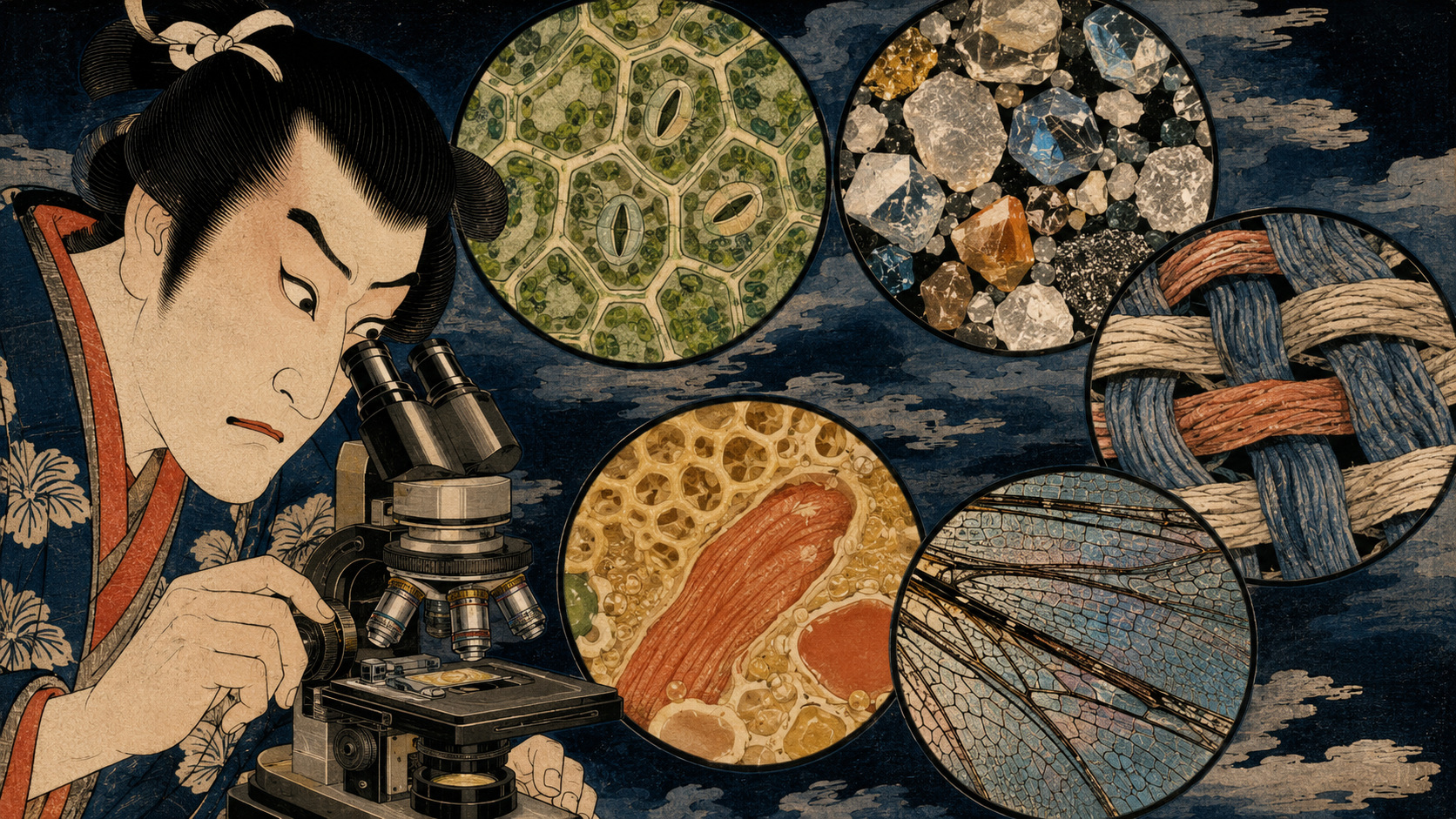
Six Doorways in the Ordinary
| Specimen | Structures to seek | A question worth carrying to the lens |
|---|---|---|
| Leaf | Veins, epidermis, hairs and—under suitable conditions—stomata | Do upper and lower surfaces, sun and shade leaves, or dry- and wet-habitat plants differ? |
| Sand | Rounded and angular grains, transparent minerals and shell fragments | How do size, roundness and color change among a river, beach and playground? |
| Cloth and paper | Weave, twisted fibers, loose ends and printed halftone dots | How do cotton, wool, synthetic fiber and washi respond to a drop of water? |
| Food | Potato starch, onion epidermis and salt or sugar crystals | What changes after heating, drying, dissolving and recrystallizing? |
| Shed insect skin or fallen wing | Hairs, scales, joints, veins and the surface of a compound eye | How does each structure serve flight, protection, movement or sensation? |
| Safely collected water | Algae, pollen, soil grains and moving microorganisms | How do surface and bottom samples, or conditions before and after rain, differ? |
Do not injure a living thing for an entry. Blood, body fluids, wastewater, mold cultures, decomposing matter and unknown hazardous specimens do not belong in a household project. Keep pond water closed, never touch it to the mouth and wash hands and tools afterward. Children should work with an adult and use commercially prepared slides when appropriate. A hobby microscope is not a safe pathogen-identification system, and shapes on a screen should never be used for medical diagnosis.
Turning One Picture into a Small Experiment
- Record the specimen, collection place, date and condition.
- Search the whole sample at low power before increasing magnification.
- Note the microscope, lens, illumination and optical magnification; record digital zoom separately.
- Where possible, calibrate against a known ruler and include a scale bar.
- Observe several areas before selecting the most attractive frame.
- Keep the original file and disclose cropping, color changes, stacking or compositing.
- Separate what was seen, what is inferred and what remains unknown.
“The leaf had holes like mouths” is already an observation. Count whether the upper and lower surfaces contain the same number. Compare a second species. If research identifies the openings as stomata, the project can move toward their role in gas exchange and water loss. The photograph stops being a conclusion and becomes an apparatus that generates the next observation.
Video has its own power. An organism in a water drop, the boundary of a growing crystal or a fiber shrinking as it dries exposes time that a still image removes. If a sequence is accelerated, reversed or assembled from selected frames, say so. Playback speed is another decision the observer has added to nature.
What Could Remain After the Contest
The prize is two microscope kits; the contest is small. Its educational value need not be measured by the number of winners. If entries were developed into a searchable archive carrying specimen, place, magnification, illumination and the observer’s own words, they could become a comparative lesson in regional sands, seasonal pollen, clothing fibers and vegetable cells. Short annotations by specialists could correct misidentification and suggest what to test next.
Citizen science requires more: common collection protocols, quality control, place and time records, expert validation and a declared research purpose. The 2026 call is not presented as that kind of study. It is an observation-and-sharing campaign. But a good observer does not rush to claim certainty. Accurately preserving what remains unknown is one of the habits from which citizen science can grow.
The World Did Not Become Smaller. It Became Wider.
Hooke drew the chambers of cork. Leeuwenhoek found moving life in a drop. Abbe defined the boundary of light, and Ruska crossed it with electrons. Japanese technicians polished the small lenses of JOICO. Modern researchers use fluorescence and computation to follow molecules. Now a child can pick up a park leaf, place a lens on a phone and send its image across Japan seconds later.
Technology has shortened the distance to observation; it has not automated the responsibility of seeing. What lies outside the focus? Is the color natural? Is this frame representative or exceptional? Has magnification been mistaken for resolution? Was a living thing harmed? Such questions turn the consumption of wonder into knowledge.
A grain of salt, the edge of cloth or a fragment of wing proves that the unknown does not require a distant planet. The contest’s best result may not be its two winning images. It may be a microscope left on someone’s desk after August 31—and a person looking again at the ordinary world, asking what to see next.
Sources and references
Entry dates, eligible work, equipment, prizes and selection criteria reflect the organizer’s July 23, 2026 announcement; applicants should confirm current terms on the official page. Product performance and distribution figures are identified as seller or developer claims and are not presented as independently verified measurements. Safety notes and the observation workflow are general editorial guidance from Japan.co.jp.
- KaraSeed: Microscopic World Observation Photo and Video Contest launch
- Official contest entry page
- microHunter official site: products, company and operating notes
- TIME: QingYing E&T iMicro C, Best Inventions 2022
- Royal Society: Hooke’s cork plate, Micrographia and the “new visible world”
- Science Museum Group: Antonie van Leeuwenhoek
- Nobel Prize: Ernst Ruska and the development of electron microscopy
- Nobel Prize: Abbe’s limit and super-resolved fluorescence microscopy
- Nikon: JOICO and CF recognized as Microscope Heritage
- Nikon: The Microscopic World exhibition at Tokyo Photographic Art Museum
- Nikon Small World: Photomicrography competition since 1975
- Nikon Small World in Motion: Video competition and judging criteria
- Prakash Lab / PLOS ONE: Foldscope origami-based paper microscope
- PubMed: EnLightenment smartphone microscopy for education and engagement
- Review of low-cost microscopes for open science
- Nature Portfolio: Image integrity and microscopy standards
- U.S. Office of Research Integrity: Best practices in scientific image processing