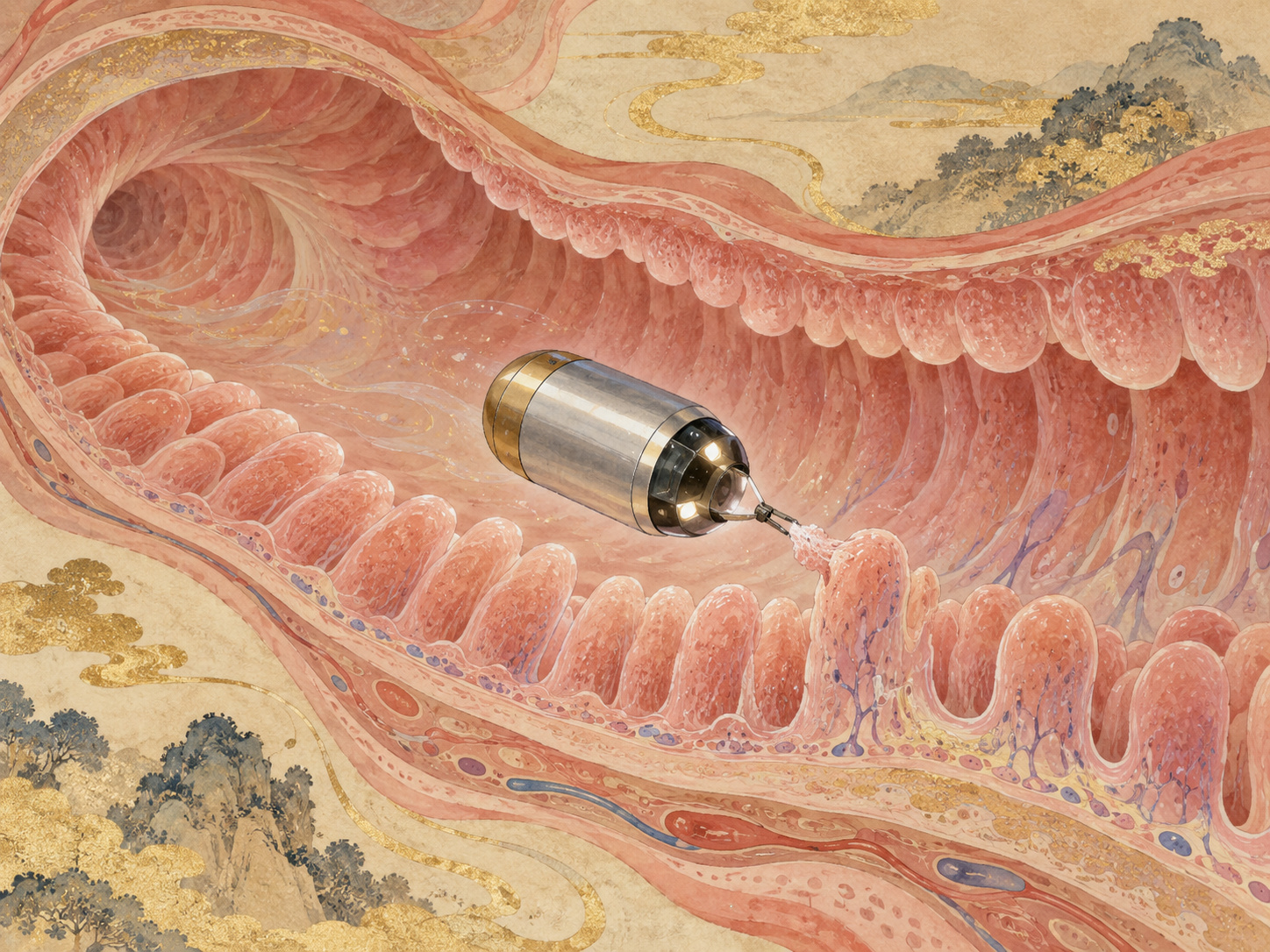
For a quarter-century, the defining achievement of capsule endoscopy has been simple to describe: swallow a camera and let it see places a conventional scope has difficulty reaching. University of Tokyo researchers are now asking a different question. What if the capsule could stop being only an observer and physically collect biological material from a chosen patch of intestinal surface?
A team led by Professor Fumihito Arai and Project Researcher Yuguo Dai at the Graduate School of Engineering has built a magnetically actuated capsule containing a retractable flexible brush and internal permanent magnets. External permanent magnets, moved by a robotic manipulator, can steer the capsule, hold it against the intestinal surface and drive the brush outward. The team demonstrated the approach using excised pig small intestine, then extracted bacterial DNA from the recovered material and successfully carried it through PCR and 16S rRNA gene sequencing.
Stool and the intestinal surface do not necessarily tell the same story
Much of what we know about the human gut microbiome comes from stool. It is easy to collect, repeatable and well suited to large genomic studies. But stool is not a uniform census of every biological niche along the gastrointestinal tract.
Microbial communities vary from stomach to small intestine to colon. They can also vary across a much shorter distance: organisms in the intestinal lumen can differ from communities associated with mucus and the epithelial surface. That surface is where microbes, nutrients, immune cells and host tissue interact most directly.
Obtaining site-specific information from that interface is harder than collecting stool. Conventional endoscopy and biopsy can access tissue, but they require substantially more equipment and clinical intervention. A swallowable device that can collect surface-associated material could occupy a different point on that spectrum.
How the brush comes out without an onboard motor
The University of Tokyo device combines a flexible brush, a screw mechanism and two internal permanent magnets. Rather than relying on a large onboard motor and battery to do mechanical work, it borrows energy and control from magnetic fields outside the body.
The researchers describe two basic movement modes. In rotational mode, rotating the external magnet causes the capsule to rotate, allowing its orientation to be adjusted. In translational mode, moving the external magnet creates an attractive magnetic force that pulls the capsule toward a target position.
Sampling uses two external magnets. One holds the capsule near the intestinal wall. The other rotates a sampling magnet inside the capsule. A screw converts that rotation into axial motion, pushing the flexible brush out of the capsule until it contacts the intestinal surface.
The brush deforms against the tissue and collects surface-associated material. Reversing the magnetic rotation retracts the brush back inside, where the sample is retained. Conceptually, the capsule becomes a remotely operated swab.
The first capsule revolution was about seeing the small bowel
Capsule endoscopy entered clinical gastroenterology in 2001 after FDA clearance and European CE marking. The engineering was remarkable for its time: a camera, illumination, battery and wireless transmitter packaged into a swallowable device.
The technology solved a real anatomical problem. Much of the small bowel had been difficult to inspect directly with conventional upper endoscopy or colonoscopy. A passive camera carried forward by peristalsis could photograph the mucosa along the way without requiring a long endoscope to traverse the entire intestine.
But passivity was also the limitation. A conventional video capsule generally goes where the intestine takes it. The physician can review what it saw, but steering it back to an interesting site, holding it still, taking a biopsy or collecting material is much more difficult.
Magnetic control turned the capsule into a robot
That limitation drove research into magnetic guidance. A magnet inside the capsule can respond to a field generated outside the body, creating force and torque without requiring a propeller or large internal motor. Robotic systems can move the external magnet in controlled trajectories.
Magnetically controlled capsule endoscopy has been developed especially for active inspection of the stomach, while research systems have explored increasingly ambitious functions: automated navigation, biopsy, local drug delivery and sample collection.
The University of Tokyo device sits in that second generation of thinking. Its novelty is not simply that the capsule can be moved magnetically. It uses magnetic control to perform a physical sampling action at the intestinal surface and then protect the collected material inside the device.
Why a brush rather than a tissue biopsy?
A biopsy is powerful because it removes tissue for histology and molecular analysis. It is also mechanically demanding. A tiny capsule must produce enough force to cut tissue, control the depth of injury, prevent excessive bleeding and securely store the specimen.
Microbiome sampling can ask a different question. If the biological target is material attached to mucus or the mucosal surface, the device may not need to cut tissue. A flexible brush can make contact, sweep up surface material and retract.
That is a meaningful design choice. It trades the depth and diagnostic richness of tissue biopsy for a potentially simpler and gentler mechanism aimed at microbial and molecular information near the surface.
The pig-intestine samples survived the full molecular workflow
The researchers tested the capsule on excised pig small intestine and found that different brush structures could recover biological material from the exposed intestinal surface.
They then asked the more important laboratory question: was the recovered material analytically useful? Bacterial DNA was extracted from capsule-collected samples, the 16S rRNA gene was amplified by PCR, and sequencing was performed.
16S rRNA sequencing is a standard approach for profiling bacterial communities because regions of the gene contain information that helps distinguish groups of bacteria. Successful sequencing does not prove that the device can diagnose disease. It does show that the engineering process—from surface contact to brush retraction to sample recovery—can preserve material usable in downstream biological analysis.
What was demonstrated — and what was not
| Function | Demonstrated | Still required |
|---|---|---|
| Capsule motion | External permanent magnets controlled rotation and translation | Stable in-vivo control through the abdominal wall and moving intestine |
| Sampling | Retractable brush contacted ex vivo intestinal surface | Quantified repeatability and site specificity in living tissue |
| Sample protection | Brush retracts into capsule after collection | In-vivo cross-contamination testing |
| Molecular analysis | DNA extraction, PCR and 16S rRNA sequencing | Clinical interpretation and broader omics validation |
| Biological model | Excised pig small intestine | Living-animal safety studies and human trials |
The hard problem is knowing exactly where the sample came from
A technology for mapping spatial microbiology needs more than a working brush. It needs location.
The small intestine is long, folded and mobile. Its shape changes with peristalsis and differs from person to person. In the laboratory, the external magnet can be precisely controlled by a robotic manipulator. In a patient, the magnetic field must act through skin, fat, muscle and other organs while avoiding excessive force on the bowel.
Even if sampling succeeds, the biological value depends on knowing whether the brush contacted duodenum, jejunum, ileum or some more precisely defined position. Future systems may need to combine magnetic localization with imaging, external tracking, transit timing or other sensors so that a biological result comes with a reliable anatomical address.
Sampling gently enough is just as important
The brush is designed to be flexible, but living intestine is not the same as excised tissue. Living mucosa has blood flow, responds to pressure and may be fragile in inflammatory disease or ulceration.
The force holding the capsule to the wall, brush extension, rotational speed, number of sweeps and contact time will all need safety limits. Existing capsule endoscopy also has a known risk of capsule retention in patients with intestinal narrowing. A capsule with moving mechanical parts adds another requirement: every component must retract reliably and remain atraumatic during transit.
The larger project: an “in-body cybernetic avatar”
The work is part of a much broader Japanese research program. Arai leads a JST Moonshot Goal 1 project titled “Structuring Spatiotemporal Environmental Information in the Body Using In-body Cybernetic Avatars,” grant JPMJMS2214.
The project envisions small devices operating at millimeter, micro and nano scales inside the body to measure variables such as temperature and pH, collect samples and eventually deliver drugs locally. JST's progress reports describe work on pill-type, capsule-type, helical-ring and stent-type platforms.
The phrase “cybernetic avatar” can sound abstract. The brush capsule makes it concrete: a small machine enters the body, is controlled from outside, travels to a target and performs a task a human operator cannot physically perform there.
1980s–1990s: Engineers develop the concept and components for swallowable wireless gastrointestinal cameras.
2001: Small-bowel capsule endoscopy enters clinical use with FDA clearance and European certification.
Late 2000s: Multiple capsule platforms emerge; research into magnetic steering expands.
2010s onward: Robotic magnetic control, AI-assisted reading and experimental biopsy/drug-delivery functions accelerate.
2022: JST selects Arai's in-body cybernetic-avatar project under Moonshot Goal 1.
2025: Yuguo Dai's doctoral work focuses on a magnetically actuated intestinal sampling capsule with a brush structure.
September 2026: The team publishes ex vivo intestinal-surface sampling with downstream 16S rRNA sequencing in Device.
This is not a replacement for stool testing
It is tempting to ask whether targeted capsule sampling could be “better” than stool analysis. There is not yet evidence to answer that question, and the comparison may be the wrong one.
Stool is valuable precisely because it is simple and repeatable. It supports longitudinal monitoring and population-scale research. A steerable sampling capsule would offer a different advantage: spatial specificity and access to material associated with the intestinal surface rather than what eventually exits the body.
The most useful future may therefore be complementary. Stool could provide a broad longitudinal signal, while targeted sampling investigates a location of interest before and after treatment or explores local host–microbe interactions that stool cannot resolve.
What has to happen before a human swallows one
The paper answers the first engineering question: can a remotely actuated capsule collect analyzable biological material from an intestinal surface? In the tested ex vivo model, yes.
Everything that follows is harder. Researchers will need to establish how much material the capsule collects, how repeatably it does so, how magnetic force changes with depth, whether the brush injures living mucosa, whether a retracted sample remains uncontaminated during transit, and whether the capsule can always pass safely through the gastrointestinal tract.
Then comes the medical question. A new sampling method has value only if the information changes understanding, diagnosis, treatment selection or monitoring. That will require comparisons with stool, intestinal fluid, biopsies and clinical outcomes in the same subjects.
Questions for the next stage
- How much mucosal injury occurs in living-animal testing?
- How much magnetic force and positioning accuracy remain through the abdominal wall?
- Can the precise sampling location be recorded and revisited?
- How effectively does brush retraction prevent cross-contamination?
- Can samples support metagenomics, metabolomics or host-biomarker analysis beyond 16S sequencing?
- How do capsule samples compare directly with stool, luminal fluid and tissue biopsy from the same subject?
- What are the retention and mechanical-failure risks in patients with intestinal narrowing?
From a camera to a machine that can make a map
The first capsule-endoscopy revolution made the small bowel visible. The next challenge is to make the gastrointestinal tract spatially measurable—not as one tube with one microbiome, but as a landscape of local environments.
Stool gives information from the exit. Biopsy can give deep information from a selected site. A steerable sampling capsule could eventually occupy a space between them: travel to a location, gently touch the surface, seal the sample away and carry it back out.
This device is not ready for patients. But it marks an important change in what a medical capsule is expected to do. It no longer has to be a camera drifting downstream. It can be a robot with a task.
If that idea survives the long path through animal studies, safety engineering and clinical validation, future gastrointestinal diagnostics may involve not just looking through the gut, but sending small machines to collect the biological geography hidden along its walls.
Sources & Reporting Notes
- The University of Tokyo Graduate School of Engineering, Sept. 30, 2026 — Primary English release describing the retractable brush, internal magnets, robotic external magnets, ex vivo pig-small-intestine testing and 16S rRNA sequencing.
- The University of Tokyo Graduate School of Engineering, Japanese primary release — Primary Japanese source for official names, terminology and technical description.
- Dai et al., Device, 2026 — Paper: 'Magnetically Actuated Brush-Structured Capsule for Intestinal Surface Sampling,' published online Sept. 29, 2026.
- Arai Laboratory publication list — Lab publication record placing the device within a broader microrobotics and biomedical-engineering program.
- University of Tokyo Mechanical Engineering dissertation record — Yuguo Dai's 2025 doctoral work on a magnetically actuated intestinal sampling capsule with a brush structure.
- JST Moonshot Goal 1: Fumihito Arai project — Primary program description for in-body cybernetic avatars and remote sensing, sampling and localized intervention.
- JST Moonshot progress report — Progress on capsule, helical-ring and stent-type in-body devices and related monitoring/sampling technologies.
- History of capsule endoscopy, PMC — Historical review of the route from early concepts to FDA clearance and CE marking in 2001.
- Systematic review of magnetically controlled capsule endoscopy, PMC — Review of magnetic steering, robotic control and expanded capsule functions.
- Gastrointestinal microbiome review, PMC — Background on biological differences between mucosa-associated and luminal/stool-associated microbial communities.
This article is based on public material available through September 30, 2026. The demonstrated experiment used excised pig small intestine. It did not establish safety or efficacy in a living animal or human. Human magnetic-positioning accuracy, mucosal-injury risk, localization of the sampling site, cross-contamination control, clinical diagnostic value and any timeline to clinical use remain unresolved.
Image rights: © 2026 Japan.co.jp. AI-assisted scientific editorial illustration. Permission is required for reuse. Contact.