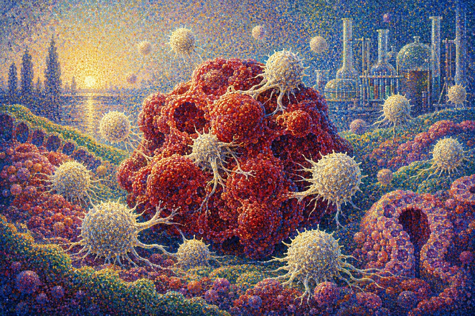
A tiny colorectal tumor is growing in a laboratory dish. It is not a flat lawn of cells. It is an organoid: a three-dimensional structure made from a patient’s tumor, preserving part of the architecture, biology and stubborn individuality of the original cancer. Researchers at Kobe University introduce gamma-delta T cells manufactured from induced pluripotent stem cells. The immune cells engage the miniature tumor and begin to damage it.
The team then moves to immunodeficient mice carrying grafts of patient-derived colorectal cancer. After the tumors take hold, the researchers deliver the cells near the tumor and, in a separate experiment designed to approximate systemic treatment of metastatic disease, through a vein. The intravenously administered cells still exert an effect at a distant tumor. In the three mice used for that experiment, tumor weights were 43% to 92% lower than in controls.
No patient was treated. The animal groups contained only three or four mice, the patient-derived tumor models came from just two people, and an immunodeficient mouse cannot reproduce a normal human immune system. Yet the result matters. Gamma-delta T cells have long offered an appealing combination of broad tumor recognition and lower dependence on patient-specific tissue matching, but their scarcity and limited expansion have frustrated mass production. Kobe’s group used iPS cells as a renewable intermediate, then tested the resulting cells against tissue that is more realistic than a conventional cancer cell line.
T cells written with a different pair of letters
Most T cells carry a receptor built from alpha and beta chains. These αβ T cells typically inspect fragments of proteins displayed by major histocompatibility complex molecules—MHC, called HLA in humans. It is an exquisitely precise system. It also helps explain why conventional T cells from one person may attack healthy tissues in another and cause graft-versus-host disease.
Gamma-delta T cells carry receptors made from gamma and delta chains. They are only a few percent of lymphocytes in human peripheral blood, but they are enriched in barrier tissues such as the intestine, skin and lungs. They can respond rapidly to signs of infection, cellular damage and metabolic stress. Their biology is more complicated than the shorthand “MHC-independent” suggests, but many γδ T-cell responses are not restricted by the classical HLA presentation system that governs αβ T cells. That makes them attractive as cells from one donor that might serve many recipients.
Immunologists recognized γδ T cells as a distinct lineage in the mid-1980s. They came to be seen as a bridge between innate immunity’s speed and adaptive immunity’s specialized receptors. They can punch pores in tumor cells with perforin, deliver cell-killing granzymes, release signaling molecules and recruit other arms of immunity. The practical obstacle has been supply. They are uncommon in blood, donors vary, and prolonged expansion outside the body can exhaust the very functions a therapy needs.
CAR-T transformed blood cancers—and exposed the solid-tumor wall
Modern cell therapy is inseparable from the rise of CAR-T treatment. A patient’s own T cells are collected, genetically equipped with a chimeric antigen receptor that recognizes a cancer marker, expanded and reinfused. Since the first U.S. approval in 2017, CAR-T products have produced durable remissions in some leukemias, lymphomas and multiple myeloma that had resisted other treatments.
But bespoke manufacturing takes time and money. A severely ill patient’s collected T cells may be few or poorly functioning, and disease can advance while the product is being made. Solid tumors add more barriers: immune cells must enter dense tissue, survive an immunosuppressive neighborhood and recognize cancers whose target markers vary from one cell to the next. In colorectal cancer, checkpoint inhibitors have been especially effective for the minority of tumors with mismatch-repair deficiency or high microsatellite instability. Most colorectal tumors still need another immune entry point.
Gamma-delta T cells may read several forms of cellular stress rather than depend on one HLA-and-antigen combination. Their anticipated lower alloreactivity supports an off-the-shelf strategy. Yet advantageous immunology does not automatically create a manufacturable medicine. Kobe’s program is an attempt to connect the two.
The 2006 revolution that made cellular rewinding possible
Induced pluripotent stem cells emerged from one of Japan’s most consequential scientific discoveries. In 2006, Shinya Yamanaka and colleagues showed that four transcription factors could return mature mouse cells to a pluripotent state. In 2007 they did so with human cells. Yamanaka and John Gurdon shared the 2012 Nobel Prize in Physiology or Medicine for demonstrating that mature cells can be reprogrammed to pluripotency.
Regenerative medicine was the most visible promise: turn iPS cells into neurons, heart muscle or retinal tissue to replace what disease had destroyed. In 2014, Kobe hosted the world’s first transplant in a clinical study using an iPS-derived retinal pigment epithelium sheet. But iPS cells can also serve as a manufacturing platform. A well-characterized starting cell can be reprogrammed, banked, expanded and repeatedly differentiated into a functional product. Japan moved iPS-derived immune cells toward patients with an iPS-NKT trial in 2020 and an iPS-derived CAR-NK ovarian-cancer trial in 2021.
The proposed Kobe therapy would not inject pluripotent cells into a patient. A donor γδ T cell is reprogrammed into an iPS cell, expanded, and then differentiated back into mature iγδT cells. Any future product would have to remove residual undifferentiated cells and pass stringent tests for identity, purity, genetic stability, potency and sterility. The iPS cell is a factory floor, not the medicine’s final form.
A three-stage research program in Kobe
The 2026 paper is the latest step in a program that has taken nearly a decade. Work first published online in 2017 and in print in 2018 established a relatively direct method to generate γδ T-cell-derived iPS cells from cultured peripheral-blood mononuclear cells without first sorting a purified γδ population. The method carried the rearranged gamma and delta T-cell receptor genes of the mature starting cells into a renewable iPS state.
In 2023, Takashi Aoi’s group reported that those iPS cells could be differentiated back into functional γδ T cells. The regenerated cells killed several kinds of cancer cell lines without strict MHC restriction and expressed perforin and granzyme B. Single-cell RNA sequencing showed that they were not merely copies of the dominant γδ T cells in blood; they resembled a rarer population with several natural-killer-like features.
The new study, led by Ryoko Futai with Aoi and colleagues, moved both manufacturing and testing closer to clinical reality. The cells were produced under feeder-free conditions, without animal support cells, and xeno-free conditions, excluding animal-derived components. Across the production process, the cells underwent roughly 80,000-fold multiplication. The team then advanced beyond familiar cell lines to patient-derived colorectal cancer organoids.
- 2017–2018: Efficient generation of human γδ T-cell-derived iPS cells.
- 2023: Regeneration of cytotoxic iγδT cells capable of killing several cancer cell lines.
- 2026: Feeder-free, xeno-free production and testing against patient-derived colorectal tumors in dishes and mice.
Why organoids raise the evidentiary bar
Established cancer cell lines are indispensable because they are consistent and easy to compare. They are also simplified. Cells selected by years of growth on plastic do not fully reproduce a patient tumor’s three-dimensional barriers, internal diversity or drug resistance.
In 2009, Toshiro Sato and colleagues reported that a single Lgr5-positive intestinal stem cell could self-organize into a crypt-and-villus-like structure and grow long-term. In 2015, researchers built a “living organoid biobank” from 20 consecutive colorectal cancer patients, showing that the cultures retained major genetic alterations and molecular subtypes while supporting drug screening. Patient-derived organoids became a bridge between cell lines and clinical trials.
They are not complete miniature patients. Most lack a full vascular system, nerves, fibroblasts, microbiota and immune environment. A sample from one region may miss other clones within the same tumor. Success against an organoid is therefore stronger evidence than success against a single cell line, but it is not proof of clinical efficacy.
What the study showed—and in what order
| Test | What it established | What remains unknown |
|---|---|---|
| Manufacturing | Reproducible feeder-free, xeno-free differentiation with roughly 80,000-fold overall expansion. | Whether GMP-scale production will retain the same yield and potency. |
| In vitro killing | Cytotoxicity against leukemia and colorectal cancer cell lines and patient-derived colorectal organoids. | Activity across many patients, genotypes, treatment histories and metastatic sites. |
| Local delivery | Tumor suppression in immunodeficient mice carrying patient-derived tumor grafts; tumor weight fell by as much as 88% in some experiments. | Persistence and efficacy inside an intact human immune and tumor microenvironment. |
| Intravenous delivery | Activity after systemic administration; the three tested mice showed tumor-weight reductions of 43% to 92%. | Distribution, dose, repeat dosing, organ toxicity and long-term persistence. |
| Normal blood cells | No clear cytotoxicity against healthy-donor peripheral-blood mononuclear cells. | Off-target effects on normal intestine, liver, lung and other tissues in humans. |
The intravenous result is particularly important. Injecting cells beside a tumor bypasses the problem of trafficking. Sending them through the bloodstream tests whether they can reach and act upon a distant target—an essential property for treating metastatic or recurrent cancer. But three animals constitute a signal for further work, not statistical certainty.
The distance between “appears safe” and “is safe”
The lack of killing against healthy-donor blood cells is encouraging. Gamma-delta T cells are expected to carry less graft-versus-host risk than conventional αβ T cells, and observations after allogeneic stem-cell transplantation support that possibility. Nevertheless, blood cells do not represent every human tissue. Cytokine release, unintended tissue recognition, trapping in the lungs or other organs, immune rejection, long-term persistence and reactions after repeated doses require separate testing.
An iPS-derived product brings additional quality questions. Residual pluripotent cells could pose a tumor-formation risk. Long culture can select genetic or chromosomal abnormalities. A product may be too weak, too strong or biologically different after freezing and thawing. Every manufacturing lot must be tested to show that a product bearing the same name truly contains the same cells and performs the same function.
Colorectal cancer is large—and profoundly diverse
Japan’s National Cancer Center reports 154,039 colorectal cancer diagnoses in 2023 and 54,416 deaths in 2024. The five-year relative survival rate is 71.4%, but that average combines disease removable at an early stage with the far harsher reality of cancer that has spread to the liver, lungs or elsewhere.
Colorectal cancer is not one enemy. Tumors differ in APC, KRAS, BRAF, TP53 and mismatch-repair status, among many other features. Even one patient’s tumor can contain multiple cell populations. Activity against organoids from two patients is encouraging, but two tumors cannot represent the disease. Researchers will need models from patients with different mutations, metastatic sites and treatment histories, especially tumors that resist checkpoint inhibitors and standard chemotherapy.
Standardized iγδT cells may also become a research instrument for understanding that diversity. The same cell product can be applied to a large panel of patient organoids. Researchers could then search for biomarkers that distinguish sensitive tumors from resistant ones, or identify combinations that break resistance without the confounding variability of a different immune-cell product for every experiment.
Why Kobe matters
The location carries unusual continuity. Yamanaka studied medicine at Kobe University before pursuing the work that led to iPS cells. The first clinical-study transplant using an iPS-derived tissue was performed in Kobe in 2014. Aoi himself worked in the broader intellectual lineage that expanded the range of cells amenable to reprogramming.
Hyogo and Kobe bring universities, hospitals, RIKEN laboratories, cell-manufacturing capabilities, regulatory expertise and biotechnology companies into close proximity. That ecosystem matters because a paper is only the beginning of a cell therapy. Clinical manufacturing, release testing, pharmacology, toxicology, trial design and long follow-up all have to work together. The new study was supported in part by Japan’s J-PEAKS university-strengthening program and the Hiroshima–Kobe–Kumamoto medical-innovation initiative.
“Off the shelf” does not mean simple or cheap
The phrase suggests a medicine waiting like a boxed product in a pharmacy. Living cells are more demanding. Even so, a well-characterized iPS master line could supply many treatment doses with more consistent starting material than patient-by-patient manufacturing. Production could occur before a particular patient needs the therapy, with cells cryopreserved and released after standardized tests. That could shorten the dangerous interval between treatment decision and infusion.
Economics will depend on more than the number of cells. Failed batches, quality testing, shipping, post-thaw viability, hospital preparation and long-term monitoring all contribute to cost. Mass production will not automatically make a therapy inexpensive. But a common batch used at many centers could improve comparability in clinical trials and make access less dependent on whether an individual patient can yield enough healthy T cells.
The next step will be decided by data, not hope
The Kobe team says it will work toward clinical-grade iγδT manufacturing and the nonclinical data package required before a trial can begin. That means testing many more patient-derived cancers and normal tissues, multiple doses, repeated administration and longer observation. The group also plans to explore lung and bladder cancers.
Another path is to add a CAR or other engineered function to the γδ T cell’s natural tumor recognition. The combination could unite broad stress sensing with precise targeting of a chosen antigen. It would also add complexity: more genetic safety testing, more manufacturing controls and a greater need to prevent excessive immune activity. A powerful cell therapy must not only attack; it must behave predictably and, where possible, be controllable.
No responsible timeline to patient availability can be inferred from the present data. Even if a first-in-human trial follows, its earliest purpose would normally be to establish dose, tolerability and safety. A dramatic percentage reduction in a mouse xenograft is not a forecast of the response rate in people.
A bridge between the dish and the ward
The achievement is best understood not as a declaration that cancer has been defeated, but as a climb to a higher rung of evidence. Gamma-delta T cells can be reprogrammed. Cytotoxic descendants can be regenerated. They can be made without animal feeder cells or animal-derived culture components. They can attack patient-derived three-dimensional cancer tissue. Delivered through the bloodstream, they can act on a distant tumor in a mouse. Each statement is limited; together they form a plausible development path.
Three histories converged in Kobe: an unconventional T-cell lineage identified in the 1980s, cellular reprogramming launched from Japan in 2006, and intestinal organoids first grown from single stem cells in 2009. In 2026, miniature patient tumors and standardized immune cells met in the same experiment. The distance to a hospital ward remains long. The advance is that researchers can now begin to measure it.
Sources and references
This report is grounded primarily in Kobe University’s release and the published study, with primary research and public sources for the histories of iPS cells, γδ T cells, CAR-T therapy and patient-derived organoids. The results are preclinical and do not establish safety or efficacy in patients.
- Kobe University: Mass-producible immune cells kill patient-derived colorectal tumors in mice (2026)
- Futai et al., Allogeneic iPSC-derived γδT cells demonstrate antitumor efficacy against patient-derived tissues, Stem Cell Reports (2026)
- Kobe University: Human iPS cells regenerated into cancer-killing γδ T cells (2023)
- Murai et al., Re-generation of cytotoxic γδT cells from human γδT-derived iPSCs (2023)
- Watanabe et al., Generation of human γδT cell-derived iPSCs (2018)
- Nobel Prize: Mature cells can be reprogrammed to become pluripotent
- Ribot, Lopes & Silva-Santos, γδ T cells in tissue physiology and surveillance
- Sato et al., Single Lgr5 stem cells build crypt-villus organoids (2009)
- van de Wetering et al., A living organoid biobank of colorectal cancer patients (2015)
- U.S. National Cancer Institute: CAR T cells and the solid-tumor challenge
- National Cancer Center Japan: Colorectal cancer statistics
- AMED: Japan’s iPS-NKT head-and-neck cancer trial (2020)
- AMED: iPS-derived CAR-NK ovarian-cancer trial (2021)
- RIKEN: First iPS-derived RPE cell-sheet transplant (2014)