A tracer signal is not a clinical diagnosis—and a possible pathway is not yet a proven one.
A single eye drop raises an unexpected question
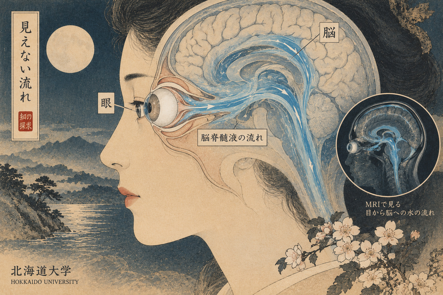
Where does the water in an eye drop go? Some drains through familiar routes on the eye’s surface. Some moves through more complicated tissues. The possibility of exchange near the optic nerve and brain has fascinated researchers, but following water molecules through a body already filled with water is unusually difficult. On October 14, the National Institutes for Quantum Science and Technology (QST) and Hokkaido University announced a specialized MRI technique that detected a labeled-water signal extending from the eye toward intracranial regions. It is a measurement advance with intriguing implications, not a new glaucoma test or a treatment for dementia.
An experiment in seeing the invisible
QST researchers including Mari Miyata and Takayuki Obata worked with Hokkaido University investigators including Professor Kohsuke Kudo and lecturer Hiroyuki Kameda. They applied specially labeled water as eye drops while acquiring MRI images. The investigators reported changes in oxygen-17 signals in the orbit, at the skull base and in regions corresponding to the brain. Those observations suggest a way to examine fluid exchange involving the eye and central nervous system. They do not, by themselves, resolve the precise route traveled by every water molecule.
Why an ordinary MRI cannot simply follow a drop
Standard clinical MRI depends primarily on signals from hydrogen nuclei, abundant in water and other molecules. It therefore provides invaluable anatomical contrast but cannot readily tell which water was just introduced. The Japanese team used water enriched with oxygen-17, a naturally rare, stable isotope detectable through tailored magnetic resonance methods. Because oxygen-17 is nonradioactive, the tracer involves no ionizing radiation. That is a specific advantage of the label, not a blanket declaration that an experimental imaging procedure has no clinical risks or constraints.
The engineering breakthrough inside the scanner
The researchers developed a dedicated radiofrequency transmit-and-receive coil for oxygen-17 and refined how its weaker signal was collected. As labeled water was repeatedly administered to participants’ eyes, the scans registered changes beyond the eye itself. This combination of tracer chemistry, hardware and image acquisition is the core technical achievement. The images are not a cinematic recording of tiny streams visibly flowing through an established anatomical pipe. Signal distribution must still be interpreted against multiple biological pathways and measurement limitations.
A longer history than the announcement suggests
This work follows years of Japanese efforts to measure water dynamics. A first-in-human study published in the Journal of Magnetic Resonance Imaging in 2023 used eye drops containing oxygen-17-labeled saline and observed intraocular changes through proton MRI. That was a different detection approach and a more restricted observation. A related manuscript posted in February 2026 on medRxiv discussed cerebrospinal-fluid signal changes after labeled eye drops, but preprints are not equivalent to peer-reviewed findings. The new October release emphasizes direct oxygen-17 MRI detection over a wider region. Understanding these steps is essential to evaluating what is genuinely new.
What the evidence does not yet establish
The joint announcement describes detected signal changes, but it does not settle the exact route from the ocular tissues to the intracranial signal. Potential pathways around the optic nerve, contributions from other distribution routes and the interpretation of the detected signal all warrant further testing. The team plans to present the work at the 54th annual meeting of the Japanese Society for Magnetic Resonance in Medicine on October 17, 2026. As of the reporting cutoff, a peer-reviewed publication specifically documenting this latest experiment was not confirmed. Conference-stage findings should be presented accordingly.
Why glaucoma gives the work urgency
Glaucoma progressively damages the optic nerve and can lead to irreversible loss of vision. Eye pressure is a key, treatable risk factor, yet cases with pressure in the conventional normal range complicate the picture. Researchers are interested in whether the fluid environment around the nerve provides additional information. This is different from claiming that eye-to-brain water exchange causes glaucoma. Nor does the new procedure demonstrate accuracy as a diagnostic test, tell clinicians when to change treatment or show that vision can be preserved.
The unsettled science of the brain’s fluid systems
A wider debate concerns how cerebrospinal fluid and interstitial fluid move through the brain, and how these processes may relate to waste clearance. Research often invokes the glymphatic system and water-channel protein aquaporin-4. Their possible roles are being studied vigorously, but the mechanisms, measurement methods and relevance to human disease remain contested in important respects. Detecting a tracer near the brain does not demonstrate removal of harmful proteins, explain Alzheimer’s disease or prove a route of therapeutic benefit.
From astronaut eyesight to brain drug delivery
Spaceflight-associated changes in eye structure and vision have heightened interest in fluid distribution in and around the head. The QST announcement points to that context as well as the future prospect of delivering treatments toward the brain through the eye. Such suggestions are research directions. The experiment used labeled water, not an active drug, and established neither drug concentration at a target nor therapeutic effectiveness or safety. Turning a tracer route into a dependable delivery system would require a separate sequence of pharmaceutical and clinical studies.
The formidable path to clinical use
Even a reproducible research image is many steps from a hospital diagnostic service. Researchers need larger samples, quantitative agreement across people and scanners, disease-versus-control comparisons, safety evaluations and evidence that an image changes decisions or outcomes. Specialized multinuclear MRI equipment is not the same as the standard setup in every radiology department. The Japanese release credits partial funding from AMED, collaboration with Siemens Healthcare K.K. and access to multinuclear MRI infrastructure. How widely and economically the method could be deployed remains unknown.
A more precise question, rather than an early miracle
The most compelling claim is also the most restrained: Japanese researchers have acquired a new kind of information about where tagged water can be detected after it enters the eye. The discovery may eventually help researchers test hypotheses that connect ophthalmology, neuroscience and medicine. Its lasting significance will depend on experimental replication and a clearer distinction between the movement of labeled water, the structure of actual flow pathways, and the mechanisms of disease. A better instrument allows better questions; answers will take longer.