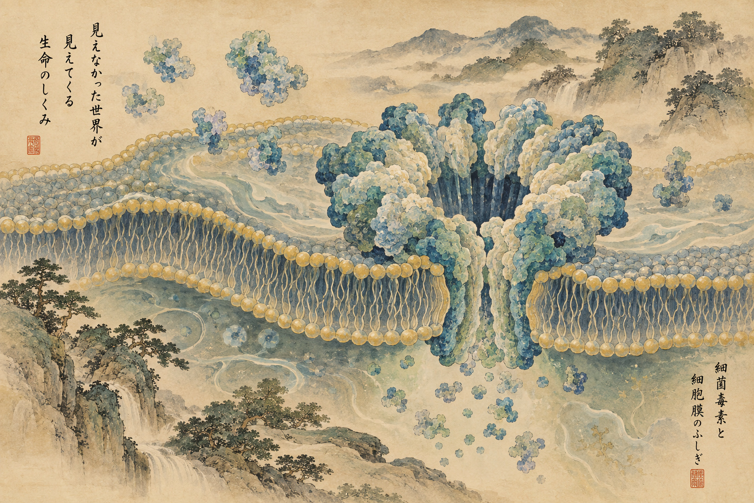
One of the most dramatic tricks in molecular biology is the conversion of a water-soluble protein into a channel that crosses a cell membrane. Cell membranes are designed to be formidable barriers. Yet a broad family of bacterial toxins can assemble on the surface, radically rearrange their own protein structure and drive a nanoscale pore through the lipid bilayer.
A collaboration between Kyoto Sangyo University and Kyoto University has now captured what happens in between. The group, led by visiting researcher Tomohito Yamada and Professor Hideaki Tsuge at Kyoto Sangyo University together with Professor Takeshi Noda and Associate Professor Yukihiko Sugita at Kyoto University’s Institute for Life and Medical Sciences, resolved multiple structural states of the membrane-binding component Ib of Clostridium perfringens iota toxin. The work appeared in the Proceedings of the National Academy of Sciences on September 28.[1]
A two-component molecular delivery machine
Iota toxin is a binary toxin. It contains an enzymatic component, Ia, and a membrane-binding component, Ib. Ib attaches to target cells, oligomerizes and forms a membrane pore. Ia then uses that pore to enter the cytosol, where it ADP-ribosylates actin and disrupts the cytoskeleton.[2]
The key is that Ib does not begin as a membrane-spanning tube. It first assembles into a ring-shaped “prepore” on or near the membrane. That assembly must then undergo a major conformational transition that produces the transmembrane pore.
Scientists have previously determined structures for prepores and completed pores. The hard part has been seeing the short-lived states between them. Intermediates are transient, structurally mixed and difficult to stabilize. Researchers could compare the starting and ending structures, but much of the actual molecular motion remained inferred.
An accidental “Ib rosette” froze the action
The key experimental opportunity came unexpectedly. Multiple Ib oligomers formed a large radial supercomplex. The researchers named it an “Ib rosette,” a term inspired by the rosette-like assemblies known from influenza hemagglutinin. Sugita and Noda said the unexpected structure became the breakthrough that exposed a phenomenon that had previously escaped observation.[3]
Inside the rosette, pore-formation intermediates that would ordinarily be fleeting were stabilized. Single-particle cryo-electron microscopy and particle classification then separated them into eight distinct prepore conformations, named Prepore I through Prepore VIII.[1]
The underlying structural data are being deposited in public databases. Prepore V, for example, is available as the seven-subunit assembly PDB 9JSL, with its corresponding electron microscopy map in EMDB as EMD-61776.[4]
The seven subunits do not transform together
A seven-membered protein ring invites a simple assumption: perhaps all seven subunits switch conformation at once, preserving symmetry throughout the process. The intermediate structures showed otherwise.
Across the series, one Ib molecule moves toward the mature conformation first. Then an adjacent subunit changes, followed by the next. The number of mature-like subunits rises one by one until the entire ring has converted. The authors describe the process as a domino-like maturation mechanism.[1]
That means the final pore can be symmetrical even though its route to that symmetry is temporarily asymmetric. It is a useful reminder that a finished molecular machine does not necessarily reveal how it was assembled.
The Φ-clamp may act as a structural checkpoint
Deep inside the Ib channel is its narrowest constriction, the Φ-clamp. It is formed by aromatic residues arranged around the pore and plays an important role when the enzymatic component Ia is threaded through the channel. Earlier structural work from the same research lineage showed the N-terminus of Ia becoming unfolded as it enters Ib and approaches this narrow constriction.[5]
In the new study, the researchers mutated an amino acid near the Φ-clamp, D452, replacing it with alanine. The Ib D452A mutant accumulated prepore-like structures instead of progressing efficiently into mature pores. That supports the idea that correct formation of the clamp region is an important structural checkpoint in the prepore-to-pore transition.[1]
“Checkpoint” here does not imply that the protein makes a decision. It describes a structural and energetic condition that appears to have to be satisfied before the next conformational step becomes favorable.
Why membrane-pore intermediates are so difficult to see
Structural biology naturally favors stable molecules. A prepore can persist long enough to purify. A completed pore can exist as a final state. The transition between them may last only briefly and represent a small fraction of particles in a sample.
Pore-forming proteins present another problem: to insert into lipid membranes they expose surfaces that interact favorably with hydrophobic membrane interiors. Outside an appropriate membrane-like environment, those same properties can make the proteins prone to aggregation. Similar sample-preparation problems have complicated decades of work on anthrax toxin, aerolysin-family toxins and other β-pore-forming proteins.[6]
How cryo-EM turned molecular motion into classifiable structures
Cryo-electron microscopy works by rapidly freezing hydrated biological samples so that water vitrifies rather than forming damaging ice crystals. Thousands or millions of particle images are then computationally aligned and combined into three-dimensional reconstructions.
The 2017 Nobel Prize in Chemistry went to Jacques Dubochet, Joachim Frank and Richard Henderson for developing cryo-EM for high-resolution structure determination of biomolecules in solution. The Nobel committee emphasized that the method can freeze biomolecules in different stages of motion and make previously unseen biological processes accessible to structural analysis.[7]
Modern single-particle analysis can also computationally separate different conformations that coexist in one dataset. That capability is exactly what makes a study with eight prepore classes possible.
A long history of trying to see a pore form
Pore-forming toxins have been structural-biology landmarks for decades. The heptameric pore of Staphylococcus aureus alpha-hemolysin, solved by X-ray crystallography in the 1990s, became a canonical picture of a β-barrel pore. Anthrax protective antigen provided another major model: it assembles as a prepore and then converts into a transmembrane channel that delivers lethal factor and edema factor into host cells.[6]
X-ray crystallography was extraordinarily powerful for stable soluble proteins and crystallizable complexes, but large membrane assemblies and mixtures of transient states were often difficult targets. The cryo-EM “resolution revolution” expanded the field from determining final pore architecture toward comparing the structures along the transition itself.
The same group had already watched the cargo begin to unfold
Tsuge’s group at Kyoto Sangyo University has spent years following the iota-toxin system. In 2020, Yamada and colleagues, working with Osaka University and others, used cryo-EM to determine the Ib pore and complexes in which Ia was bound to it. They showed that when Ia engages the pore, part of its N-terminal alpha helix loses its folded structure and extends toward the constriction inside Ib.[5]
In 2022, the group helped determine analogous complexes from the binary CDT toxin of Clostridioides difficile, capturing early structural events as its enzymatic component begins to unfold at the translocation pore.[8]
The new work moves one step earlier in time. Instead of asking how a toxin enzyme passes through a completed pore, it asks how that pore itself comes into existence.
Do not confuse iota toxin with ordinary food-poisoning mechanisms
Clostridium perfringens causes a range of human and animal diseases, but different strains carry different toxin repertoires. Common human C. perfringens food poisoning is primarily associated with enterotoxin CPE, whereas iota toxin is classically associated with type E strains. The contribution of iota toxin to human disease is considerably less established than that of several other major C. perfringens toxins.[9]
The significance of the present study is therefore better understood as a fundamental discovery about pore-forming proteins than as a direct explanation of the usual mechanism of human C. perfringens food poisoning.
Pores are biological weapons—and useful nanomachines
Pore-forming proteins are not limited to bacterial toxins. Related membrane-perforating mechanisms appear in immunity and programmed cell death. Across biology, the ability to convert a soluble protein into a membrane-spanning assembly is a recurring strategy for killing cells, moving molecules or controlling membranes.[6]
Engineers have also learned to exploit nanoscale pores. Nanopore sensing can detect DNA, RNA and proteins as individual molecules alter ionic current through a tiny channel. The more precisely researchers understand pore diameter, charge, hydrophobicity, gating and assembly, the more control they may eventually gain over synthetic pores and molecular transport systems.
The Kyoto researchers explicitly identify artificial nanopores and intracellular molecular delivery as possible long-term areas where a structural understanding of pore maturation could become useful.[1]
That does not make this a near-term drug or delivery device
The work is structural and mechanistic. It does not demonstrate a therapy that blocks D452, an antibiotic against iota toxin, or a functioning drug-delivery device based on Ib.
There is another limitation. The Ib rosette was experimentally valuable precisely because it stabilized states that are normally transient. Whether the eight classes occur with the same lifetimes and populations on a living cell membrane remains a separate kinetic question. Structural snapshots must eventually be connected to real-time measurements, membrane composition and cellular physiology.
But the central discovery survives those caveats: the missing middle of the transition is populated by real structural states, and those states show the conformational change propagating around the ring rather than occurring simultaneously.
Structural biology is moving from portraits toward storyboards
For much of its history, structural biology excelled at producing portraits of molecules: the stable state before a reaction, the stable state after it. Biology itself is dynamic. Proteins open, rotate, bind, unfold and cross membranes.
Eight prepore structures are not a movie. But ordered correctly, they become something closer to a molecular storyboard—a sequence showing how one architecture transforms into another.
Seven Ib proteins form a ring. One changes first. Then its neighbor. Then the next. Eventually a symmetrical pore emerges from an asymmetric journey. At the scale of a few billionths of a meter, the bacterial machine appears to build itself not with a single synchronized snap, but one molecular domino at a time.
Sources
- Kyoto University / Kyoto Sangyo University detailed research release, Sept. 30, 2026
- Clostridium perfringens Iota-Toxin: Structure and Function
- Kyoto University research news, Sept. 30, 2026
- RCSB PDB 9JSL: iota toxin Ib, Prepore V
- Kyoto Sangyo University, cryo-EM analysis of iota-toxin translocation, 2020
- Advances in cryoEM and its impact on β-pore forming proteins
- The Nobel Prize in Chemistry 2017 — cryo-electron microscopy
- Kyoto Sangyo University, structural analysis of C. difficile binary toxin CDT, 2022
- Towards an understanding of the role of Clostridium perfringens toxins in human and animal disease
- Institute for Life and Medical Sciences, Kyoto University, Oct. 1, 2026