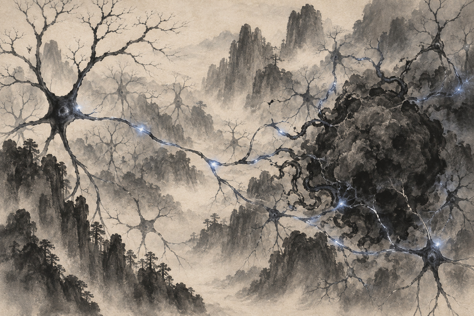
The brain is not merely the place where a glioma happens to grow. It is an electrically active ecosystem that a tumor can enter, listen to and exploit. For a decade, the emerging field of cancer neuroscience has found increasingly direct ways in which neuronal activity accelerates glioma. The new study from Japan adds the other pedal: a neural circuit capable of applying a brake.
Researchers at Nagoya City University, Institute of Science Tokyo, the Japanese Foundation for Cancer Research, Teikyo University, the University of Tokyo, Hokkaido University, Nagoya University, Fujita Health University and the National Hospital Organization Osaka National Hospital report that adult glioma cells receive functional synaptic input from inhibitory neurons. Their paper, “Inhibitory Circuits Restrain Adult Glioma Progression by Suppressing Calcium-Driven Oncogenic Programs,” appeared in Neuron on September 7 in the United States, September 8 in Japan.
The corresponding authors are Naofumi Uesaka, Reo Maruyama and Daisuke Kawauchi. Uesaka, Kohei Kumegawa and Takaki Watanabe are listed as co-first authors. The work combines anatomy, electrophysiology, manipulation of neural activity, calcium imaging and molecular analysis to move beyond the observation that neurons and tumor cells sit close together.
The tumor is wired into the brain
Gliomas are primary tumors arising from cells of the brain and spinal cord. Glioblastoma, one of the most aggressive adult forms, infiltrates normal tissue beyond the visible mass, making complete removal extraordinarily difficult. Treatment generally combines as much safe surgery as possible with radiotherapy and temozolomide. Japan’s 2024 clinical guideline cites a median survival of 20.0 months among patients receiving standard therapy and a five-year survival rate of about 16% in national registry data.
Against that clinical background, the new work asks a basic biological question: what messages does a glioma receive from the organ it invades? Electron microscopy revealed structures consistent with synaptic contacts from inhibitory neurons to tumor cells. Electrophysiological recordings then showed that those contacts worked—that they transmitted signals and reduced electrical activity in the glioma cells.
When the researchers increased inhibitory-neuron activity in mouse brains, calcium activity within the tumors became weaker and less widespread. Tumor-cell proliferation fell and the mice survived longer. Reducing inhibitory-neuron activity produced the opposite result: proliferation increased. Those bidirectional interventions make the case stronger than a static correlation between a neural marker and tumor size.
A causal chain through calcium
Calcium ions do much more than build bone. Inside a cell, changes in calcium concentration carry information, converting an external stimulus into commands affecting gene expression, metabolism, movement and division. In glioma, coordinated calcium transients can connect neural input to collective tumor behavior.
The team tested whether calcium was simply associated with the inhibitory effect or carried it. Directly lowering calcium activity in tumor cells, without first stimulating inhibitory neurons, was enough to suppress growth and extend mouse survival. Artificially restoring calcium activity weakened the tumor-suppressing effect of inhibitory circuitry. Together, the interventions support a sequence: inhibitory synaptic input lowers tumor calcium activity, and lower calcium activity restrains progression.
Molecular analysis placed YAP1 and mTOR downstream. Both sit within programs that help cells interpret mechanical, nutrient and growth signals and sustain proliferation or survival. When tumor calcium activity fell, YAP1- and mTOR-related signaling also declined. Growth suppression was reproduced in patient-derived glioma models, an important step beyond a single laboratory cell line.
Patient-derived, however, does not mean patient-tested. Cells grown or transplanted for an experiment retain selected features of a human tumor but do not reproduce the full person: immunity, circulation, normal neural function, drug metabolism and treatment toxicity. No dose, device, approved drug or clinical protocol follows directly from this paper.
| Question | What the study establishes | What remains unknown |
|---|---|---|
| Connection | Functional inhibitory synapses onto adult-glioma cells | How consistently they occur across patients and tumor classes |
| Causality | Inhibitory activity and lower tumor calcium restrain growth and extend mouse survival | Whether any intervention improves human survival |
| Mechanism | Reduced calcium activity and weaker YAP1/mTOR programs | The safest tumor-selective target and therapeutic window |
| Translation | Growth suppression in patient-derived models | A validated drug, device or clinical trial |
How an accelerator became a two-pedal system
The importance of the finding becomes clearer against the field’s short history. In 2015, Humsa Venkatesh and colleagues reported in Cell that active neurons release neuroligin-3, or NLGN3, which promotes high-grade glioma growth and activates PI3K–mTOR signaling. It was powerful evidence that the brain’s normal activity could be converted into a tumor-supporting signal.
Two Nature papers in 2019 went further. They demonstrated bona fide excitatory synapses between neurons and glioma cells, involving AMPA-type glutamate receptors. The tumors were not passive neighbors: they integrated electrical and calcium signals into invasive, proliferative behavior. A cancer had become electrically wired into the organ it was destroying.
The Japanese study does not overturn those discoveries. It changes the circuit diagram. Excitatory synapses and activity-dependent growth factors can press the accelerator, while inhibitory input can restrain adult-glioma calcium programs. The translational question becomes more precise than “Can neural activity be stopped?” It becomes: can tumor-promoting circuitry be weakened while a protective circuit is preserved or exploited?
2005 — Radiotherapy plus temozolomide changes the standard of care for glioblastoma.
2015 — Activity-dependent NLGN3 secretion is shown to promote glioma growth.
2019 — Excitatory neuron-to-glioma synapses are demonstrated in landmark studies.
2025 — GABAergic input is reported to promote pediatric diffuse midline glioma.
2026 — The Japanese team identifies an inhibitory circuit that restrains adult glioma.
Why “inhibitory” does not always mean anti-tumor
The word “inhibitory” invites a dangerously easy conclusion. GABA, the brain’s principal inhibitory neurotransmitter, does not reduce electrical activity in every cell under every condition. Its effect depends on ion gradients across the cell membrane, receptor composition, developmental state, tumor subtype and anatomical location.
In 2025, Tara Barron and colleagues reported a seemingly opposite result in pediatric-type diffuse midline glioma. GABAergic neuron-to-glioma synapses depolarized those tumor cells and promoted proliferation, in part because their chloride handling made the signal excitatory rather than inhibitory. That is not necessarily a contradiction. It is evidence that different gliomas translate the same neurotransmitter into different biological instructions.
That contrast places a hard boundary around the new claim. “GABA suppresses brain cancer” would be inaccurate. It would also be unsafe to infer that sedatives, anti-seizure medicines or other GABA-active drugs treat glioma. Such medicines act across normal circuits governing awareness, memory, movement, breathing and seizures. Before translation, researchers must identify which inhibitory-neuron subtype connects to which adult tumor, through which receptor and ion environment, in which patients.
Five gates between discovery and treatment
- Measure variation by molecular subtype, grade, brain region, prior treatment and patient.
- Map which tumor cells receive inhibitory input and whether resistant cells escape it.
- Find a tumor-selective target that leaves cognition, movement and seizure control intact.
- Test interactions with surgery, radiotherapy, temozolomide and tumor-treating fields.
- Replicate independently, complete safety studies and only then design human trials.
One route may be to avoid changing whole-brain activity and instead intervene at a tumor-side junction: calcium regulation, the receptor receiving the input, or the signaling link to YAP1 and mTOR. That could, in principle, be more selective. Yet these pathways also serve essential functions in normal tissue. Naming a target is not the same thing as producing a safe medicine.
The paper’s strongest contribution is therefore not a proposed prescription. It is an experimentally tested chain of events linking synaptic input, electrical state, calcium, oncogenic programs, proliferation and survival. Each link becomes a measurable point at which future studies can ask why one patient’s tumor responds and another’s does not.
The unit of cancer biology is changing
Cancer was once framed largely as a collection of mutated cells. Immunology expanded that frame to include immune systems; vascular biology added blood supply and metabolic exchange. Cancer neuroscience now asks investigators to treat a brain tumor as a disease embedded in a functioning circuit. The genome still matters, but so do the neurons a tumor contacts and the timing and chemistry of the signals it receives.
If researchers can see only the accelerator, the obvious ambition is to disconnect the tumor. Finding a brake suggests a subtler goal: shift the local circuit balance toward restraint. The 2025 pediatric result warns that this balance will not be universal. What opened this week is not a shortcut to treatment, but a more exact map of the disease—and a demanding new path toward precision cancer neuroscience.
- Nagoya City University, “Discovery of a neural circuit that suppresses brain tumors,” September 8, 2026 — institutions, experiments, authors and stated limitations.
- Uesaka et al., Neuron, 2026, doi:10.1016/j.neuron.2026.08.008 — original study and bibliographic record.
- National Cancer Center Japan, Cancer Information Service: glioma — disease and treatment background.
- Japan Society for Neuro-Oncology, 2024 glioblastoma guideline — standard care and Japanese outcomes.
- Venkatesh et al., Cell, 2015 — activity-dependent NLGN3 and glioma growth.
- Venkataramani et al., Nature, 2019, and Venkatesh et al., Nature, 2019 — excitatory neuron-to-glioma synapses.
- Barron et al., Nature, 2025 — growth-promoting GABAergic input in diffuse midline glioma.
Editorial note: Japanese institutional names, titles, paper terminology and author names were checked against Japanese primary sources. Japan.co.jp has not inferred unpublished effect sizes or a patient benefit. The broad wording “brain tumors” in the supplied headline is narrowed throughout the article to the adult-glioma models actually studied.