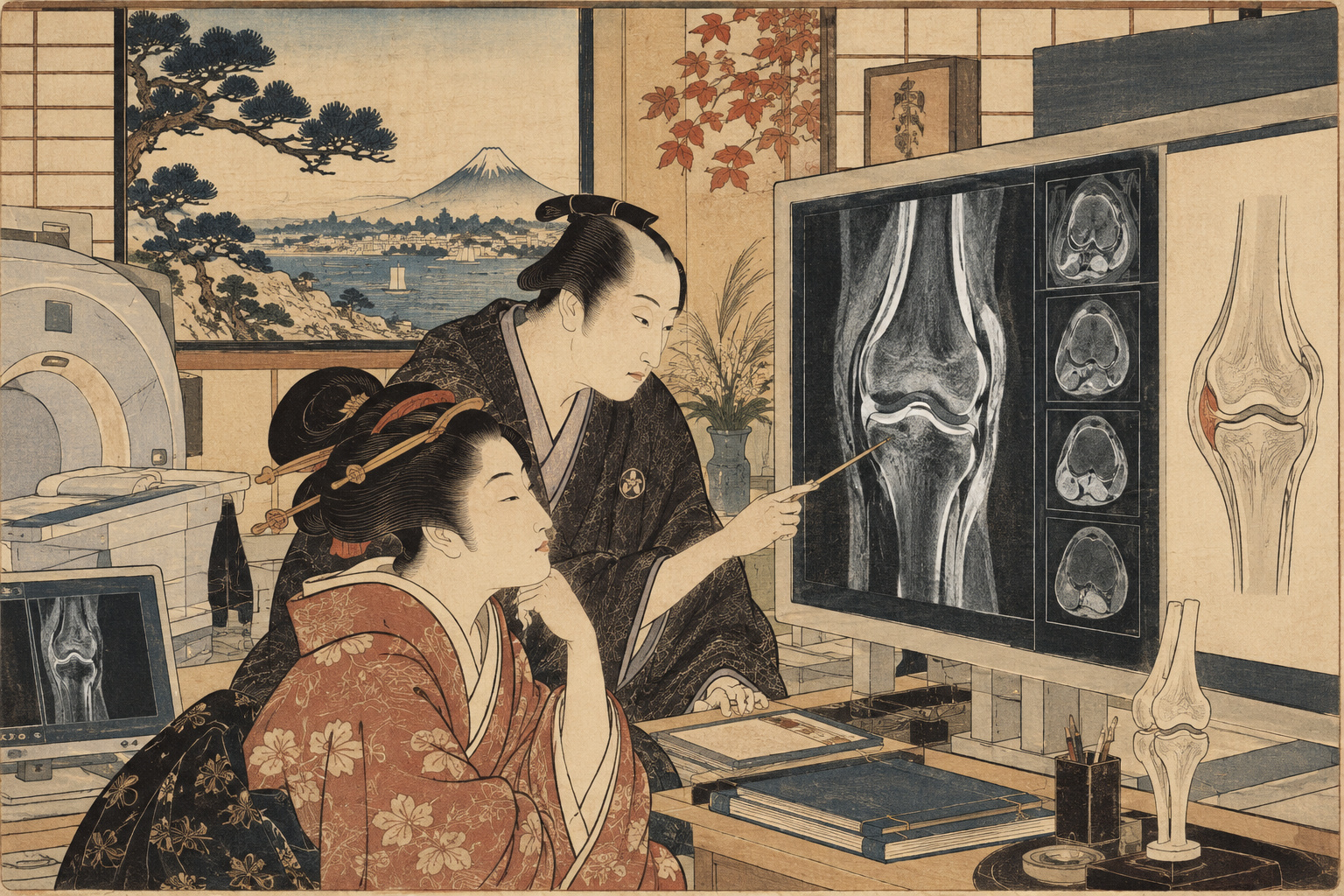
A knee does not become arthritic on the day it starts to hurt. Cartilage can thin, the meniscus can shift, ligaments can change and bone can remodel long before symptoms become severe. Modern MRI can see many of those changes. That creates a second, harder problem: deciding which abnormalities are warnings and which are merely things that happen to be visible.
On September 15, Institute of Science Tokyo announced a new phase of the Kanagawa Knee Study with Kanagawa Prefecture and Fujifilm. Ichiro Sekiya and colleagues at the Center for Stem Cell and Regenerative Medicine will re-examine people who took part in the original project about eight years ago, linking their earlier MRI, radiographs and lifestyle information to new imaging, questionnaires and clinical outcomes. The aim is to test whether structural clues present before major symptoms can predict later knee pain and osteoarthritis progression. No new general population cohort is being recruited for this follow-up. [1][2]
One clue has a deliberately simple name: the pocket sign. On MRI it appears as a small fluid-equivalent signal at the interface where the meniscotibial ligament attaches near the medial tibial cortex. In a June 2026 Scientific Reports paper, the sign became more common as radiographic osteoarthritis severity increased. It was already present in 39% of knees graded KL 0—knees without radiographic osteoarthritis. [3]
That sounds like an early marker. It is not yet a predictive test. The published study was cross-sectional: it took a snapshot of different knees at one time. It could show association with severity, but it could not establish whether the pocket appeared before degeneration, alongside it, or after it. The new follow-up is designed to answer precisely that temporal question.
The June study associated it with radiographic severity, but the authors explicitly said the temporal sequence could not be inferred. The eight-year follow-up is an observational test of prediction, not a recommendation to send asymptomatic adults for routine knee MRI. [3]
A tiny fluid signal at a load-bearing junction
The medial meniscus is a crescent of fibrocartilage that spreads load across the inside of the knee. The meniscotibial ligament helps anchor its outer edge to the tibial plateau. If that restraint weakens, the meniscus may move outward—a change called medial meniscus extrusion, or MME. An extruded meniscus covers less of the cartilage it is supposed to protect, altering how force passes through the joint.
In the 2026 study, Hisako Katano, Nobutake Ozeki, Yusuke Nakagawa, Hideyuki Koga, Sekiya and colleagues defined the pocket sign conservatively: a definite fluid-equivalent signal directly touching the medial tibial cortex at the ligament-bone interface. To reduce false positives from a single image artifact, the signal had to continue at the same anatomical location across at least three adjacent reconstructed MRI sections. [3]
The wording matters. The authors treated the sign as a surrogate imaging marker potentially related to meniscotibial ligament detachment, not as direct proof that the ligament had detached. In six knees examined during total knee arthroplasty, the surgeons found insertion-site changes that supported the MRI interpretation. Six operative observations are useful corroboration, but they are not a large pathological validation series. [3]
The first study found a gradient, not a forecast
The pocket-sign analysis began with 573 Kanagawa Knee Study participants. After withdrawals and ineligible data, 559 were screened; 90 with lateral knee osteoarthritis were excluded, leaving 469 people. Their median age was 54, with a range of 30 to 79 years, and the sex distribution was close to even. The research intentionally excluded people with a history of knee OA, lower-extremity trauma, previous knee surgery, rheumatoid arthritis or prolonged continuous hospital visits, helping the cohort capture structural changes outside a typical advanced-arthritis clinic. [3]
Pocket prevalence rose from 39% in KL grade 0 to 100% in KL grade 4. In a multivariable model, each one-grade increase in Kellgren-Lawrence severity was associated with 2.56 times the odds of having a pocket. Sex and BMI were not significant predictors in that model; the age association was weak and inconsistent when examined within KL strata. [3]
Among the 120 KL0 knees with a pocket, the researchers identified 128 separate pockets because eight knees had two. Sixty-three percent were located anterior to the medial collateral ligament, suggesting a non-random anatomical pattern. Meniscal extrusion at those sites was generally mild—again consistent with the idea that this may be a change that can exist before obvious radiographic disease. [3]
But a marker found in roughly two out of five radiographically normal knees cannot be useful simply because it is visible. Prediction requires the next set of numbers: how many pocket-positive people progress, how many remain stable, and how many pocket-negative people develop disease anyway.
The follow-up is built to connect past images with future outcomes
The registered Kanagawa Knee Study Follow-up is observational, with a target sample size of 560. The anticipated start date is September 24, 2026, and the registry lists follow-up through March 31, 2028. The team will link baseline information to new non-contrast MRI, knee radiographs, body measurements and questionnaires obtained about eight years later. There is no treatment allocation and no intervention in clinical management. [4][5]
The outcomes are concrete. Structural progression includes at least a one-grade increase in radiographic KL classification. Symptom change will be assessed with the Knee injury and Osteoarthritis Outcome Score, or KOOS. Clinical progression includes reaching right-knee total knee replacement or osteotomy. Baseline explanatory variables include 3D MRI measurements, meniscal extrusion and meniscotibial pocket morphology. [4]
2018 — The Kanagawa Knee Study begins collecting 3D MRI and radiographic data.
2020 — The group reports automated 3D MRI segmentation and links meniscal coverage with cartilage measurements.
2022–2024 — Studies map cartilage defects, meniscal extrusion and the relationship between X-ray grades and 3D MRI structure.
June 2026 — The pocket-sign cross-sectional study appears in Scientific Reports.
September 2026 — The roughly eight-year follow-up launches.
March 2028 — Registered end of follow-up.
X-rays are not obsolete; they answer a different question
The story can be misread as “MRI replaces X-ray.” That is not what the researchers or Japanese guidelines say. Weight-bearing radiographs remain a practical foundation of knee OA diagnosis and severity grading. The Kellgren-Lawrence system, developed from mid-20th-century radiographic work, scores osteophytes and joint-space narrowing from 0 to 4 and remains widely used in epidemiology and clinical studies. [6]
MRI adds tissues that X-rays cannot directly show well: cartilage, menisci, ligaments, synovium, marrow lesions and fluid. Japan’s 2023 knee OA guideline says MRI and ultrasound can contribute to diagnosis, clinical evaluation and progression assessment because changes occur across the whole joint from early stages. [6]
Yet more sensitivity creates a paradox. The same guideline cites research in which 89% of middle-aged or older people with KL0 radiographs had at least one MRI abnormality. If almost everyone has something detectable, detection alone is not a diagnosis of impending disability. [6]
The challenge is no longer simply to make hidden knee changes visible. It is to identify which visible changes carry useful information about the future.
Why this cohort keeps returning to the meniscus
The Kanagawa Knee Study has spent years building a structural story around the meniscus rather than treating osteoarthritis as cartilage wear alone. A 2020 paper described fully automated 3D MRI software that segmented cartilage and meniscus with high accuracy and used data from 561 volunteers to quantify the relationship between meniscal coverage and cartilage measurements. [7]
A 2022 analysis of women in their 70s found tibial cartilage defects extending in contact with the medial meniscus rather than appearing at random locations away from it. In 2023, the group linked AI-determined KL grade to medial meniscus extrusion and cartilage thickness. A 2026 cohort paper reported medial meniscal tears in 18.2% of 469 participants, with tear prevalence rising sharply with KL grade and extrusion. [8][9][10]
Taken together, the program is testing a broader mechanical hypothesis: if the meniscus loses position or attachment, load shifts, cartilage becomes exposed, and structural OA may progress along that path. The pocket sign is attractive because it may capture one very early component of that sequence.
Turning a one-day reconstruction into a five-minute workflow
Longitudinal imaging studies only work if image processing can scale. Science Tokyo says the group’s 3D knee MRI analysis, developed with Fujifilm and implemented through SYNAPSE VINCENT, can display bone, cartilage and meniscus in three dimensions and quantify features such as cartilage thickness and volume. Work that once required roughly a day to construct a single 3D case can now be produced from the dedicated MRI dataset in about five minutes. [1]
That speed is not just a convenience. It is what makes repeated quantitative analysis across hundreds of participants feasible. The follow-up will also combine imaging with symptoms, lifestyle, physical function and Kanagawa’s ME-BYO health indicators, acknowledging that pain and disability cannot be read from anatomy alone. [1]
Early detection is only useful if something can follow it
There is no established drug that reliably restores the damaged structure of an osteoarthritic knee. The Kanagawa announcement explicitly notes that medicines can relieve pain but that a drug proven to reverse damaged joint structure has not been established. Japanese treatment guidelines still place education, exercise, weight management where appropriate, physical therapy and symptom-directed medication at the center of conservative care. [2][6]
So even if the pocket sign proves predictive, it will not automatically create a one-step “positive scan, take this drug” pathway. Researchers would still need to determine whether knowing the sign changes behavior or treatment in ways that reduce symptoms, slow structural progression or delay surgery.
A useful risk model may ultimately combine imaging with age, muscle strength, alignment, previous injury, weight and other structural features. The value of an eight-year cohort is that it can test combinations against actual outcomes instead of assuming that one image feature carries the whole story.
A cohort designed for early change also has limits
Science Tokyo describes the original group as including many Kanagawa Prefectural Government employees and retirees, particularly people with desk-based work and without prolonged treatment for knee disease. That is a strength when the aim is to see changes before a hospital population becomes severely symptomatic. [1]
It also limits generalizability. People with physically demanding jobs, elite sports histories, major obesity, previous injuries or established knee disease may follow different structural pathways. Even a successful eight-year prediction model would need external validation in other Japanese populations and, eventually, outside Japan.
The Fujifilm role is part of the evidence trail
The new follow-up is a collaboration among Science Tokyo, Kanagawa Prefecture and Fujifilm. The university says it will use Fujifilm’s image-analysis application under a joint research agreement and receive technical advice. In the pocket-sign paper, one co-author is identified as a Fujifilm employee and Sekiya disclosed research funding from the company. [1][3]
Those ties do not invalidate the work; imaging research often depends on industry software and engineering. They do make transparency, preregistration, reproducible methods, peer review and independent replication especially important as a research marker moves toward possible clinical use.
Japan has millions of radiographic knees and far fewer painful ones
The Japanese Orthopaedic Association guideline cites estimates of roughly 25 million people with radiographic knee OA changes and about 8 million with symptoms such as pain, stiffness or swelling. Estimates vary by definition and source, but the gap illustrates a basic problem: structural change and lived disease overlap imperfectly. [6]
That is why the right public-health goal is not mass MRI simply because MRI can find more. It is risk stratification—finding a pattern specific enough to identify people who are genuinely more likely to deteriorate while avoiding medicalizing the many abnormalities that never become clinically important.
Can a small pocket seen eight years ago tell a useful story now?
The pocket sign is visually intuitive: a little fluid-like gap where a meniscus-supporting ligament meets bone. But the scientific question is demanding. Did people who had that sign eight years ago develop more extrusion, more cartilage loss, worse KOOS scores or more surgery than those who did not?
If the answer is no, the sign may remain an interesting correlate of current anatomy. If the answer is yes—and if the result holds outside this cohort—it could become one piece of a much earlier way to define knee OA risk.
The most important result will not be that MRI can find osteoarthritis before an X-ray. It will be whether an MRI finding can distinguish the knee that is merely different from the knee that is actually heading toward disease.
Sources and references
- Institute of Science Tokyo: Capturing knee changes before pain with MRI — Kanagawa Knee Study follow-up announcement, September 15, 2026
- Kanagawa Prefecture: Kanagawa Knee Study follow-up announcement, September 15, 2026
- Katano H, Ozeki N, Nakagawa Y, et al. Meniscotibial ligament pockets on MRI are associated with radiographic severity of medial knee osteoarthritis. Scientific Reports. 2026.
- UMIN-ICDS: Kanagawa Knee Study Follow-up Study, UMIN000062772
- Japan clinical research portal: Kanagawa Knee Study Follow-up Study
- Japanese Orthopaedic Association: Clinical Practice Guideline for Knee Osteoarthritis 2023
- Aoki H, et al. Relationship between medial meniscus extrusion and cartilage measurements in the knee by fully automatic three-dimensional MRI analysis. BMC Musculoskeletal Disorders. 2020.
- Katano H, et al. Three-dimensional MRI shows cartilage defect extension with no separation from the meniscus in women in their 70s with knee osteoarthritis. Scientific Reports. 2022.
- Sekiya I, et al. Association of AI-determined Kellgren-Lawrence grade with medial meniscus extrusion and cartilage thickness by AI-based 3D MRI analysis in early knee osteoarthritis. Scientific Reports. 2023.
- Medial meniscus tears in early-stage medial knee osteoarthritis: Prevalence and type in a Japanese cohort. Journal of Orthopaedic Science. 2026.
- Institute of Science Tokyo Center for Stem Cell and Regenerative Medicine: Kanagawa Knee Study publication summaries
- Institute of Science Tokyo: Kanagawa Knee Study Follow-up 2026 participant information
Evidence reviewed September 16, 2026, Japan time. The pocket sign is not yet an established predictor of future knee OA. The published evidence is cross-sectional; the newly launched longitudinal study is designed to test predictive value. Statements about screening, prevention and implementation are Japan.co.jp analysis unless otherwise attributed.