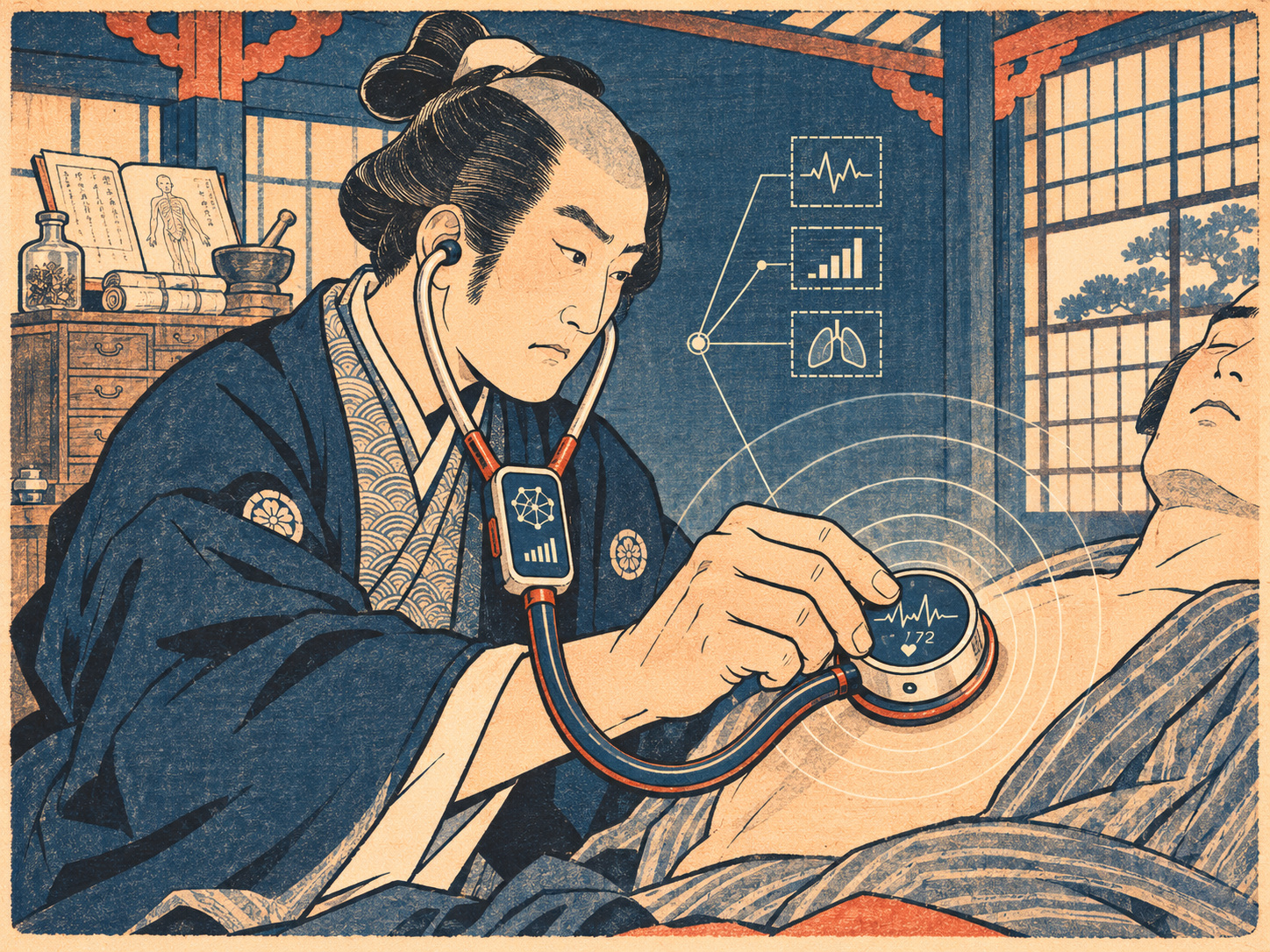
- A familiar instrument, a different kind of question
- What exactly is the condition being detected?
- Two independent groups, not a single demonstration
- The four numbers readers should not confuse
- Did AI really beat seven cardiologists?
- The architecture behind an eight-second recording
- The 200-year argument about listening
- From AI electrocardiograms to AI auscultation
- Approval belongs to the device, not the new algorithm
- What would real clinical usefulness look like?
A familiar instrument, a different kind of question
The stethoscope has long been a symbol of the physician’s bedside judgment. Yet a heart can sound unremarkable even when its principal pumping chamber is no longer working as it should. A Japanese research team is testing whether a digital recording of the sounds, combined with a brief electrical trace, can reveal a risk that the unaided ear may miss.
Juntendo University announced on November 12, 2026, that a team including graduate researcher Eiichiro Sato, Associate Professor Nobuyuki Kagiyama, Professor Tohru Minamino and AMI Inc. had developed an artificial-intelligence model that had identified reduced left ventricular pumping function in an independent multicenter group with an area under the ROC curve of 0.904. The paper appeared online in NEJM AI on October 15. The university announcement is new this week; the underlying journal publication is older.
What exactly is the condition being detected?
The left ventricle pumps oxygenated blood to the body. Clinicians often quantify its performance by left ventricular ejection fraction, or LVEF: the proportion of blood expelled from that chamber with each contraction. The Japanese study defined left ventricular systolic dysfunction as an LVEF of 40% or less on echocardiography.
That is not a diagnosis of every form of heart failure. Patients may have heart failure despite preserved ejection fraction, and reduced ejection fraction may exist before obvious symptoms emerge. The model was trained to flag a particular echocardiographic finding, not to replace a full assessment of breathlessness, swelling, medical history and other tests.
Two independent groups, not a single demonstration
The prospective study recruited adults who already had a clinical reason to undergo an echocardiogram. Using AMI Inc.’s Cardio-EGG phonocardiography system, the researchers recorded heart sounds and a single-lead electrocardiogram simultaneously at four conventional chest locations, eight seconds at each site.
The development cohort comprised 2,359 people, averaging 69.7 years of age; 8.9% met the low-LVEF definition. A separate registry at six other centers supplied an external-validation cohort of 964, averaging 68.6 years; 7.6% met the definition. By testing on people and hospitals outside the development group, the researchers went beyond reporting how well an algorithm fit its original sample.
The four numbers readers should not confuse
The independent validation produced an AUROC of 0.904, with a reported 95% confidence interval from 0.876 to 0.930. That figure summarizes the model’s discrimination across possible thresholds. It does not mean that 90.4% of individual diagnoses were correct.
At the operating point reported in the paper, sensitivity was 86.3%, specificity 74.2% and overall accuracy 75.1%. The university also reported a negative predictive value of 98.5% in its validation sample. Each measures something different: the ability to detect reduced LVEF when present, avoid false alarms when absent, make correct decisions overall, and estimate the chance of not having the condition after a negative result. Predictive values shift when the condition’s prevalence changes.
Did AI really beat seven cardiologists?
In a reader substudy involving 100 selected patient recordings, seven cardiologists assessed the heart sounds. AI achieved 82.0% accuracy against 62.0% for a majority-vote cardiologist consensus, a statistically significant difference (P=0.004). It is a noteworthy but tightly defined comparison.
The algorithm had access to both acoustic and electrocardiographic information; the physicians were assessing sounds. The substudy was not a 964-patient contest, nor a test of physicians using their full clinical resources. In real practice a cardiologist can review medical history, an ECG, laboratory results and echocardiography. The evidence supports a claim about a specific listening task—not that machines are generally better cardiologists.
The architecture behind an eight-second recording
The instrument simultaneously captures the vibrations produced by cardiac activity and a single view of the heart’s electrical rhythm. The research combined a transformer-based neural network, useful for relationships across a time sequence, with a one-dimensional convolutional network that can extract local features from waveform patterns.
The combined four-site model outperformed sound-only and ECG-only versions reported by the university (AUROCs of 0.832 and 0.804, respectively). A simpler two-location configuration retained an external-validation AUROC of 0.903 in the journal report. That is potentially valuable for workflow, although a reduced number of recording sites cannot automatically be assumed equivalent under every clinical condition.
The 200-year argument about listening
In 1816, René Laennec began using a tube to listen to sounds within the chest in Paris. His 1819 treatise transformed listening into a teachable diagnostic method, linking characteristic sounds with pathological findings. The original wooden, single-ear instrument was a far cry from the familiar twin-ear stethoscope, which developed later.
The central idea endured because sound offers information without a major procedure. Electronics eventually allowed auscultation to be recorded, replayed and measured. This study represents another step: rather than asking a clinician simply to recognize a murmur or an extra sound, researchers paired sound with ultrasound-defined heart function, searching for predictive signals hidden in the waveform. That remains a statistical inference, not a direct acoustic measurement of ejection fraction.
From AI electrocardiograms to AI auscultation
The concept of using AI to find low cardiac ejection fraction predates this paper. In a 2021 randomized pragmatic study involving primary-care teams across 45 clinics or hospitals, Mayo Clinic researchers reported that giving clinicians AI-enabled ECG results increased new diagnoses of low ejection fraction within 90 days, from 1.6% under usual care to 2.1% in the intervention group.
The Juntendo-led research asks a related but different question: can two inexpensive types of waveform, obtained during one encounter, identify which patients referred for imaging have impaired systolic function? One cannot infer from either study that the same approach will automatically work in all communities, devices or languages. The most persuasive evidence of benefit will come from prospective deployment studies measuring what patients actually gain.
Approval belongs to the device, not the new algorithm
An important regulatory distinction appears in the Japanese announcement. AMI’s AMI-SSS01-series Cardio-EGG recording system has medical-device approval, the university says. The artificial-intelligence model evaluated in this study remains a research model and has not received medical-device approval.
The research was conducted with AMI, supported by the company and a Japan Society for the Promotion of Science grant (JP25K19371). The published paper lists AMI employees among the authors and describes independent study auditing and monitoring. These disclosures belong alongside the results so that readers can judge how the evidence was produced.
What would real clinical usefulness look like?
This was not a random sample of people at a community health screening. Participants were already scheduled for an echocardiogram because of a clinical indication, and their prevalence of reduced LVEF reflects that selection. Sensitivity, specificity, predictive values, acquisition quality and workflow may differ at a rural clinic, a noisy emergency department or an asymptomatic screening event.
A useful next trial would ask not only whether the model scores highly in an analysis of prospectively collected recordings, but whether integrating it into care brings forward needed echocardiograms, reduces missed dysfunction without overwhelming specialists, and ultimately improves treatment and health outcomes. It would also test fairness across ages, cardiac rhythms, other diseases and different equipment.
The achievement is substantial because it was evaluated prospectively across institutions. But the remaining distance is equally concrete. In the two centuries since Laennec, the question has moved from what the physician can hear to what a machine can detect. The answer that matters next is whether either can help more people receive the right care in time.
Sources and verification
The Japanese university announcement and journal record were checked. No original patient interviews or clinical replication were conducted.