- A notorious protein has a second job
- The brain’s border is not just a wall
- A loss-of-function experiment in human vessel cells
- The surprising involvement of BACE1
- Another signal: nitric oxide
- What the mice added—and did not prove
- From James Parkinson to the synuclein era
- The dilemma for therapies that lower alpha-synuclein
- What remains unresolved
- A more complete picture of what protects the brain
A notorious protein has a second job
Alpha-synuclein is best known for what can go wrong. When it misfolds and accumulates in abnormal assemblies, it is implicated in Parkinson’s disease and related disorders. But researchers in Japan have identified a strikingly different role for the same protein: in tiny blood vessels of the brain, it appears to help keep a protective barrier intact.
Published in the peer-reviewed journal Aging Cell on November 9, 2026, the study examined human brain microvascular endothelial cells and mice. Removing or reducing alpha-synuclein disrupted the junctions between vascular cells and made an experimental barrier more permeable. The finding challenges a simple idea that the less alpha-synuclein there is, the better.
That does not mean the protein has been declared harmless—or that a Parkinson’s treatment has been discovered. It means location, form and physiological function matter. A protein associated with disease-causing aggregates can also perform useful work in healthy tissues.
The brain’s border is not just a wall
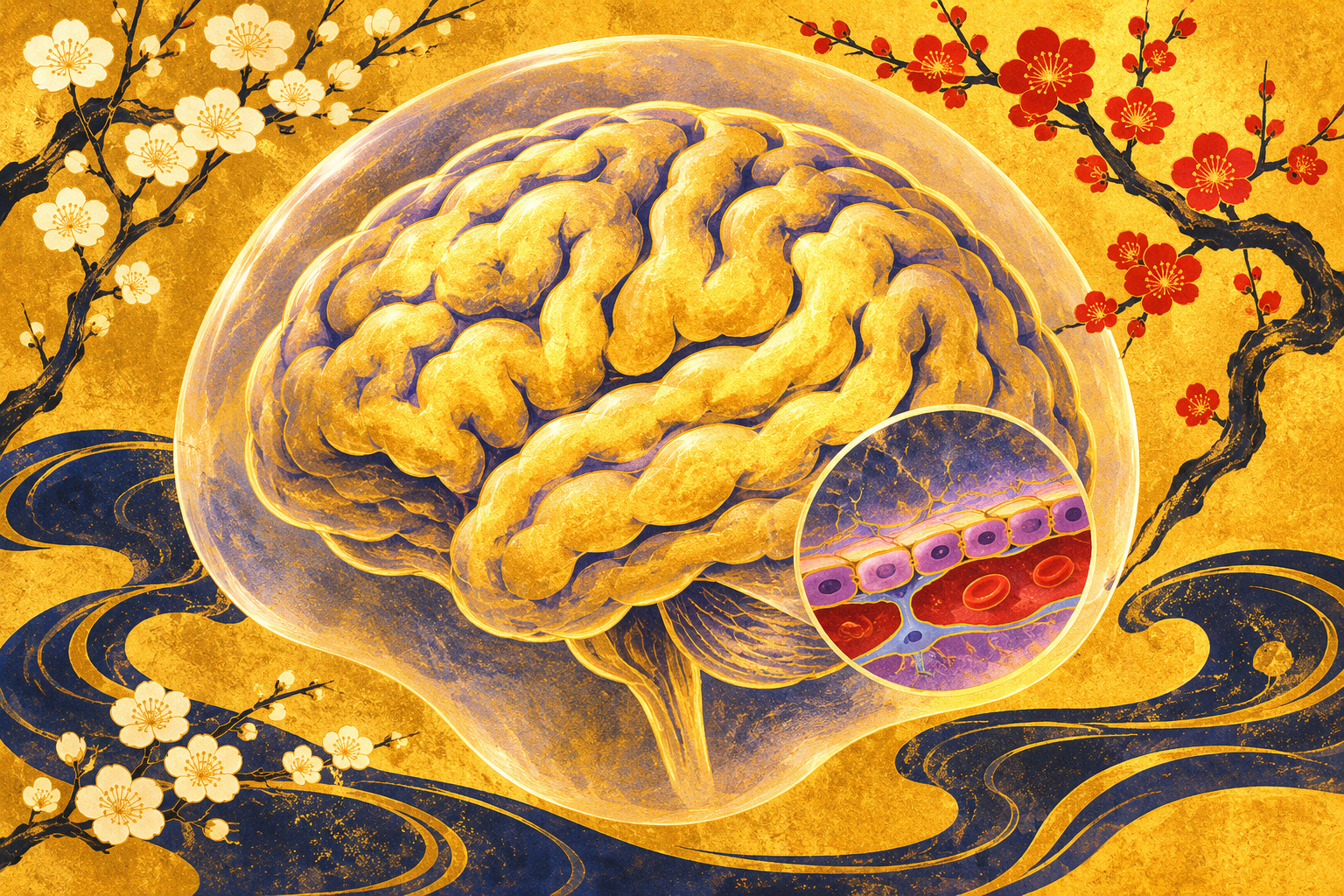
Every moment, the brain needs oxygen and nutrients delivered by blood. It also needs to limit exposure to many circulating substances. The blood–brain barrier is the selective interface that makes both possible. Specialized endothelial cells line microscopic vessels and form tight junctions, while supporting cells and transport systems help regulate exchange.
The barrier is neither impermeable nor a single plastic-like membrane. It is a living system that admits necessary molecules, removes waste and responds to changes in the vascular environment. When it becomes less selective, proteins and inflammatory signals that ordinarily remain in the blood may gain greater access to brain tissue.
Researchers have long documented changes in barrier function during aging. The open question is which molecular failures in the vascular cells make the interface more vulnerable—and whether those failures can be separated from other age-related changes.
A loss-of-function experiment in human vessel cells
The researchers reduced expression of SNCA, the gene that produces alpha-synuclein, in cultured human brain microvascular endothelial cells. They observed poorer proliferation and migration, reductions in tight-junction proteins including occludin and ZO-1, and increased permeability across the cell layer.
The result is important because this is not a study of neurons accumulating toxic aggregates. It is an investigation of endothelial cells losing a normally present protein. The same molecule can participate in different biological processes in different cell types.
A cell-culture experiment is not a miniature version of an aging patient. Gene-silencing procedures, growth conditions and the absence of an intact brain all limit direct clinical interpretation. The study used additional techniques and animal experiments to build a more persuasive mechanistic case.
The surprising involvement of BACE1
The loss of alpha-synuclein coincided with increased expression and activity of BACE1, an enzyme familiar to Alzheimer’s researchers for its role in generating amyloid-beta peptides. Here, the relevant question was what the enzyme was doing within endothelial cells and their junctions.
When the team suppressed both SNCA and BACE1, tight-junction proteins and experimental barrier performance recovered. This rescue strengthens the argument that BACE1 contributes to the damage downstream of alpha-synuclein loss. It is a mechanistic clue, not proof that an existing BACE1-targeting medicine will protect human brains.
BACE1 has physiological functions, and a laboratory intervention does not establish an acceptable dose, safety profile or benefit in people. Drug discovery must reckon with the entire organism, not only a repaired cell layer.
Another signal: nitric oxide
Using transcriptomic and proteomic techniques, the investigators identified disruption across pathways related to nitric oxide signaling, including SIRT1–eNOS and the NO–sGC–cGMP axis. Nitric oxide is not merely a chemical associated with pollution; inside the body it helps regulate blood-vessel signaling.
The research suggests that weakened signaling may contribute to BACE1 upregulation and endothelial stress. A nitric-oxide donor partially reversed some experimental changes, offering additional support for the proposed pathway without settling every causal step.
Vascular aging involves many interacting processes—metabolism, inflammation, supporting cells and tissue-specific stress. This paper identifies a plausible circuit within that larger network rather than claiming to explain all blood–brain barrier aging.
What the mice added—and did not prove
Mice engineered to lack SNCA across the body exhibited leakage of blood-borne material in several brain regions, reduced tight-junction markers, and signs of microvascular cellular stress. The researchers also found lower alpha-synuclein in isolated brain microvessels of normally aged mice.
Additional cell experiments suggested that SNCA-depleted endothelial layers were more vulnerable to amyloid-beta42 oligomers under conditions that left control layers relatively unaffected. This supports a vulnerability hypothesis; it is not evidence that the researchers reproduced Alzheimer’s disease in people.
A global knockout affects many tissues, making it difficult to isolate every effect to endothelial cells. Future work will need cell-specific models and carefully characterized human samples to determine how much of this pathway operates in ordinary human aging and neurodegenerative disease.
From James Parkinson to the synuclein era
In 1817, the British physician James Parkinson described a distinctive movement disorder in An Essay on the Shaking Palsy. In the twentieth century, the discovery of dopamine deficits and the development of levodopa transformed the understanding and symptomatic treatment of Parkinson’s disease.
The story took another turn in 1997, when genetic findings connected SNCA with familial Parkinson’s disease and research identified alpha-synuclein as a major component of Lewy bodies. Misfolding and aggregation consequently became central themes in the field.
But a molecule discovered in diseased tissue also has a normal biology. The new endothelial study illustrates why understanding that biology can be as important as documenting the pathological deposits.
The dilemma for therapies that lower alpha-synuclein
Some experimental strategies seek to reduce the production, propagation or harmful aggregation of alpha-synuclein. The new findings raise a question about tissue specificity: might it be important to preserve normal endothelial function while tackling toxic forms of the protein?
It would be premature to infer that any particular treatment in development is unsafe. Different interventions target different molecular forms, reach different organs and produce different degrees of suppression. The study did not evaluate patients receiving a synuclein-directed therapy.
The practical lesson for researchers is to measure off-target and vascular effects thoughtfully. The lesson for patients is equally clear: no change to prescribed Parkinson’s medication follows from these laboratory results.
What remains unresolved
The strongest claims are about the experimental systems: SNCA depletion reduced junctional integrity in human brain endothelial cell cultures; BACE1 co-silencing rescued measured defects; and mouse observations were consistent with a role for endothelial alpha-synuclein in barrier maintenance.
The paper does not show that reduced endothelial alpha-synuclein causes Parkinson’s disease in people, nor that boosting it prevents dementia or restores cognition. No clinical trial was conducted. Human tissue studies, cell-specific models and longitudinal evidence remain important next steps.
Independent work also identifies other contributors to age-associated barrier leakiness, including altered transport across endothelial cells. These mechanisms may coexist. A persuasive new pathway is not a universal explanation.
A more complete picture of what protects the brain
Parkinson’s research often focuses on the neurons that produce dopamine. Yet those neurons depend on vessels that deliver nutrients and maintain a finely regulated chemical environment. The aging brain is an ecosystem of neurons, vascular cells, immune activity and supporting tissue.
The Japanese team’s contribution is not a reversal of the evidence linking abnormal alpha-synuclein to neurological disease. It is an expansion of the question: what happens when the normally functioning protein is missing from a very different cell type?
The answer is still incomplete, but it may reshape how researchers define a useful therapeutic target. The challenge is no longer merely to remove a troublesome molecule. It is to understand which forms are harmful, which functions are protective, and how to distinguish them without damaging the system researchers hope to save.
Sources and verification method
Reporting method: Independent reading of the peer-reviewed Aging Cell paper (published Nov. 9, 2026; DOI 10.1111/acel.70770), the Japanese institutional announcement and historical sources. No original researcher interview or patient trial was undertaken.
- University of Tokyo Hospital / collaborating universities — Japanese press release, November 9, 2026
- Wang et al., Aging Cell (2026), DOI 10.1111/acel.70770
- Aging Cell, volume 25 issue 11 — research abstract and publication date
- OmicsDI / jPOST dataset PXD085451 — supporting proteomics data
- Parkinson’s Foundation — historical research milestones
- Parkinson’s Foundation — explanation of alpha-synuclein biomarkers
- Neuron (2026) — independent research on barrier deterioration in aging
- Brain (2026) — research on erythrocyte-derived alpha-synuclein and the BBB